119
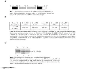
A. Fig A. : Domain structure organisation of LmPDI analysed using CDD database. a and a’ represents the catalytic domains (CGHC); b-b’ the non catalytic domains; and c, the acidic terminal extension with KDEL ER localization signal. B. 1. 26. 119. 145. 219. 231. 333. 355. 454. 477. a.

119
E N D
Presentation Transcript
A Fig A. : Domain structure organisation of LmPDI analysed using CDD database. a and a’ represents the catalytic domains (CGHC); b-b’ the non catalytic domains; and c, the acidic terminal extension with KDEL ER localization signal B 1 26 119 145 219 231 333 355 454 477 a b b’ a’ c Table B: Amino-acids alignment analysis between L. major PDI (LmPDI; Tritrypdb ID: LmjF36.6940) and their orthologues from various Leishmania species, L. donovani PDI (L. don PDI; GeneBank ID: CBZ39163.1), L. infantum (L. inf PDI; Tritrypdb ID: LinJ36_V3.7280), L. mexicana (L. mex PDI; GeneBank ID: CBZ26559.1), L. amazonensis (L. ama PDI; GeneBank ID: ABJ98154.1) and L. braziliensis (L. braz PDI; Tritrypdb ID:LbrM35_V2.7320). PDI sequences were compared using BLAST programs from NCBI and Tritrypdb databases. Alignment results are expressed in terms of identities (Id) and similarities (Sim) percentages. C R P A }LmPDI (52kDa) 1 2 3 Fig C: Expression levels of native LmPDI in Promastigotes and Amastigotes. (A) 5.106 metacyclic promastigotes and amastigotes of L. major HV clone (lanes 2 and 3) and 5µg of purified recombinant LmPDI (lane 1) were analyzed by Western blotting. The blot was immunostained with the rabbit anti-LmPDI antiserum (lanes 1, 2 and 3). Brackets indicate the native LmPDI doublet recognized by the polyclonal antibody. Supplemental data 2