Nanoparticle Optics Part 1
Nanoparticle Optics Part 1. Gold and Silver Nanoparticles Group 1 – Luke, Matt, and Jeff. Theory.

Nanoparticle Optics Part 1
E N D
Presentation Transcript
Nanoparticle Optics Part 1 Gold and Silver Nanoparticles Group 1 – Luke, Matt, and Jeff
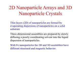
Theory • The color of the sol arises from a combination of absorption and scattering of light and depends on particle size. More specifically it is due to a resonance of the free electrons in the metal particle. The light’s electromagnetic field causes them to slosh back and forth (plasmon oscillations). • At a characteristic frequency which depends of the size and the metal, the sloshing is the most intense. This is the frequency where plasmon oscillations are excited. The plasmon resonance is easily seen in the extinction spectrum of the sol. • The particles experience the constant buffeting of Brownian motion which also helps to keep them in suspension.
Objective • Learn about scattering and absorption in gold and silver nanoparticles. • Visually observe how particle size effects scattering. • Learn to use a Tyndall beam. • Observe sub-diffraction limit nanoparticles in the optical microscope. • Become familiar with optical and electron microscopy.
Procedure: Gold Nanoparticles • Bring to a boil 50 mL of 2.5×10⁻⁴ M chloroauric acid solution • Add 0.16 mL of 34 mM sodium citrate solution to the boiling solution while stirring • After a minute will be faint blue and then darkening over 5 min to a brilliant red • Repeat procedure with 0.30 mL and 1.0 mL of sodium citrate to produce three samples of different sized gold nanoparticles. • Fill cuvette with sample and place in spectrophotometer. Record absorbance of each sample from 200 nm to 800 nm.
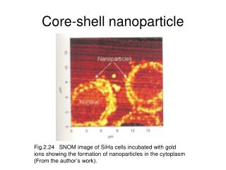
Procedure: Optical Microscopy • Use micropipette to place 20 mL of sample on a microscope slide. • Place cover slip over sample. • Place slide under 20X objective lens for total magnification of about 200X. • Use coarse and fine focus knobs to find nanoparticles. Use the plane of cover slip and slide as a reference. • Observe stationary nanoparticles on cover slip and slide. Nanoparticles in solution are randomly moving because of Brownian motion.
Procedure: Electron Microscopy • Using a micropipette, put a few drops of sample on light side of carbon grid. • Wick away excess with filter paper. • Let sample dry for several hours. • View sample in TEM and take photos for later analysis.
Results: Visual Inspection • Solution changed from blue to red within five minutes as expected. • Sample with 0.16 mL sodium citrate turned a duller red than the other two samples.
Results: Absorption Spectra • Fit of Mie Plot data to Spectrophotometer data resulted in gold nanoparticle radii of 14.8 nm, 15.6 nm, and 35 nm (diameter of 29.6 nm, 31.2 nm, and 70 nm). • Shoulder on 0.3 mL sample is because of larger particles also in the solution.
Results: TEM 0.16 mL, 100 nm scale 0.16 mL, 100 nm scale 0.3 mL, 20 nm scale
Results: TEM 1 mL, diffraction 0.3 mL, 100 nm scale 0.3 mL, 20 nm scale
Analysis: TEM • Particles in each picture were measured with a ruler to get a size distribution. • Data agrees well with estimations from absorption spectra
Analysis: TEM Diffraction • Measured radii of diffraction rings to determine lattice constant of gold.
Questions • Approximate Size: r = 14.8 nm, 15.6 nm, and 35 nm • Atoms per nanoparticle • For r = 14.8 nm: • For r = 15.6 nm: 9.4 * 10^6 atoms/nanoparticle • For r = 35 nm: 106 * 10^6 atoms/nanoparticle
Questions • Fraction of atoms on surface • For r = 14.8 nm: • For r = 15.6 nm: 0.0033 • For r = 35 nm: 0.0015
Questions • Number of nanoparticles per mL • For r = 14.8 nm: • For r = 15.6 nm: 1.6 * 10^9 nanoparticles/mL • For r = 35 nm: 0.14 * 10^9 nanoparticles/mL
Questions • Nanoparticle surface area per mL • For r = 14.8 nm: • For r = 15.6 nm: 0.049 cm^2/mL • For r = 35 nm: 0.022 cm^2/mL