Download

1 / 9
90 likes | 129 Views
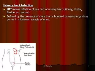
Explore the anatomy and function of the lower urinary tract including ureters, bladder, and urethra. Learn about transitional epithelial cells, urothelium, and differences between male and female urethras. Discover the mechanisms that allow the tract to adapt to varying volumes and protect against urine effects.

E N D
URINARY TRACT – III The lower urinary tract Maria M. Picken MD, PhD mpicken@luc.edu
Outline: III – the structure and function of the lower urinary tact ureters urinary bladder urethra
Objectives: General objectives: - to identify the main structures, function and location - to analyze the relationship between microscopic structure and function Specific objectives: • On H&E stained section of the ureter, identify the urothelium, muscularis, adventitia • On H&E stained section of the urinary bladder, identify the urothelium, detrusor muscle • Summarize the function of the transitional epithelial cells of the bladder • Contrast and compare the male versus the female urethra
E L M Ureter: L-lumen, E-epithelium (urothelium) M-muscularis a conduit to drain urine at low pressures from the kidney to the bladder http://www.meddean.luc.edu/lumen/MedEd/urology/abnurtdv.htm
Trigone – no rugae (ridges) Serosa (peritoneum) upper & sides transitional epithelium (urothelium) lamina propria with discontinuous muscularis mucosae detrusor muscle, aka deep muscle
The urothelium (aka transitional epithelium): • renal pelvis, ureters, bladder, parts of urethra • 3-5 cell layers, which can contract and expand The term transitional epithelium does not imply that this epithelium is in actual transition from one type to another, but rather refers to the appearance of the cells, which changes as the organs with which they are associated are distended versus not distended: • cuboidal when bladder is not distended • flat when bladder is distended Umbrella cells non-distended (relaxed) bladder distended bladder
The transitional epithelium 1. accommodate the fluctuation in volume 2. protect against the caustic effects of urine - a powerful barrier to urine Umbrella cells: • apical exocytosis of specialized fusiform vesicles (aka discoid vesicles) during distension of the bladder provides additional fragments (reserve) of cell membrane, which are incorporated into the cell membranes, allowing them to stretch in a full bladder 2. the apical plasma membrane of umbrella cells, facing the urine, is covered with rigid-looking plaques, which, together with tight junctions, form a specialized membrane compartment that represents one of the tightest and most impermeable barriers in the body Urothelial carcinoma: bladder, pelvis, ureter, urethra
Female versus male urethra • Epithelium: • transitional near bladder • squamous near external orifice • mucus urethral glands • Pathology: • urine cytology Male urethra - connected with the reproductive system
Questions? mpicken@luc.edu