Download
1 / 55
550 likes | 775 Views
UCONN NMRbox Summer Workshop 2016 Chemical Shifts, Protein Structure, and Multivariate Analysis frank.delaglio@n ist.gov. Frank Delaglio. June 21 2016 Version 3. Identify many H-H short range NOE distances Supplement with torsions from J-Coupling values Assume standard peptide geometry

E N D
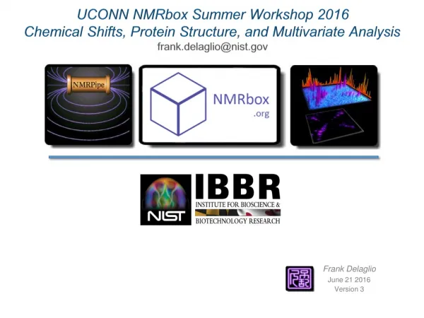
UCONN NMRbox Summer Workshop 2016 Chemical Shifts, Protein Structure, and Multivariate Analysis frank.delaglio@nist.gov Frank Delaglio June 21 2016 Version 3
Identify many H-H short range NOE distances Supplement with torsions from J-Coupling values Assume standard peptide geometry Use simulated annealing to find a structure which matches distances
Identify many H-H short range NOE distances Supplement with torsions from J-Coupling values Assume standard peptide geometry Use simulated annealing to find a structure which matches distances The network of distances is complicated, likewise the NOE spectra are complicated NOE distances are only qualitative A given peak might be the only evidence of an interaction A mis-assigned peak can be similarly problematic
Alternative Protein Structural Data from NMR F F Y Beta Sheet Y Alpha Helix Protein Folds Can Be Characterized by F,Y Backbone Angles F delaglio@nmrscience.com
Subtract Residue-Specific Random Coil Shift to form Secondary Shift
Chemical Shift and Backbone Structure Motif Match database triplet with target, based on sum-of squares difference in chemical shifts, plus residue type homology term. Use central residue as predictor of phi and psi.
The TALOS-N database contains 580 proteins. On average, TALOS-N makes consistent predictions for about 90% of the residues. About 3.5% of the unambiguous predictions made by TALOS-N differ from the crystal structure. On average, the RMSD as reported by TALOS-N for the consensus predictions was 8.7 degrees for φ, and 8.5 degrees for ψ. The actual RMSD of the "correct" predictions relative to the crystal structures was 12.3 degrees for φ, and 12.1 degrees for ψ (which includes the uncertainty in the X-ray derived angles). TALOS-N can identify the chemical shift signature of a given χ1 rotamer for about 50% of the residues, all corresponding to cases where no extensive rotamer averaging is taking place. http://spin.niddk.nih.gov/bax/nmrserver/talosn Y Shen and A Bax: Protein backbone and sidechain torsion angles predicted from NMR chemical shifts using artificial neural networks. J. Biomol. NMR, 56, 227-241 (2013).
SPARTA+ http://spin.niddk.nih.gov/bax/nmrserver/sparta Y Shen and A Bax: SPARTA+: a modest improvement in empirical NMR chemical shift prediction by means of an artificial neural network. J. Biomol. NMR, 48, 13-22 (2010).
Consistent blind protein structure generation from NMR chemical shift data • ProcNatlAcadSci USA, (2008) 105, 4685-4690 • Yang Shen • Oliver Lange • Frank Delaglio • Paolo Rossi • James M. Aramini • Gaohua Liu • Alexander Eletsky • Yibing Wu • Kiran K. Singarapu • Alexander Lemak • AlexandrIgnatchenko • Cheryl H. Arrowsmith • Thomas Szyperski • Gaetano T. Montelione • David Baker • Ad Bax
Using SPARTA Chemical Shift Prediction to Improve ROSETTA Scoring Function
0.57 0.64 0.60 0.70 0.66 1.10 2.03 2.07 0.69
Structures of Two Designed Proteins with HighSequence Identity NMR structures vscsRosetta models NMR structures of Ga88 and Gb88 Mean-to-mean backbone RMSD 1.31A Patrick A. Alexander, Yanan He, Yihong Chen, John Orban, and Philip N. Bryan PNAS, 2007, 104:11963-11968 PNAS, 2008, 105:14412-14417 1.07A
CS-Rosetta Structure NMR Structure https://csrosetta.bmrb.wisc.edu/csrosetta?key=6106ea029d9c
NMR Spectral Fingerprinting of Biologic Therapeutics Using Interferograms: Exploiting Non-Uniform Sampling Without the Need for Spectral Reconstruction Yves Aubin Luke Arbogast Robert Brinson John Marino • 2D HN/N correlated NMR to reveal High Order Structure • Practical 2D H/C correlated Methyl NMR of mAbs at natural abundance • Multivariate analysis for easy evaluation of NMR fingerprint data • NMR spectral fingerprinting without spectra Goal: Use NMR to provide direct answers about properties such as protein fold, excipient effects, glycosylation, stability, and aggregation. Changes in these properties can reduce the efficacy of a biotherapeutic, or cause harmful immune responses. Strategy: Use 2H, 13C, and 15N isotopic labeling to guide the development of natural abundance methods. frank.delaglio@nist.gov
Fourier transforms are used to convert time-domain data to frequency-domain, and the information content is similar in all domains. Time-Domain Interferogram Frequency Domain = =
Non-Uniform Sampling (NUS) Can Reduce Measurement Time, but Also Requires Special Reconstruction Methods Conventional FID Interferogram Fourier Spectrum NUS FID NUS Interferogram NUS Fourier Spectrum
Methods Such as IST Can Produce Excellent NUS Spectra … Conventional FID Interferogram Fourier Spectrum NUS IST Reconstruction NUS FID NUS Interferogram
Methods Such as IST Produce Excellent NUS Spectra, but these Non-Linear Methods Can Also Change Lineshape and Apparent Noise Distribution FT vs NUS FT vs FT Scaled Intensity Scaled Intensity Scaled Intensity
Top 10 Drugs by US Sales Accounts for $60 Billion of $315 Billion Four of the Top 10 are Biologics Biologics are life-changing and life-saving therapeutics. They are also expensive, an issue for everyone in the healthcare system. As originator biologics go off patent, less expensive biosimilars can be produced. Development and manufacture requires monitoring high-order structure, aggregation, stability, and modifications such as glycosylation. Statistics from IMS Health Inc. as quoted on medscape.com, May 6, 2015
Most Drugs work by Targeting a Specific Protein or Class of Proteins Most familiar drugs are small organic molecules, often discovered by synthetic chemists making many variations of a molecular scaffold. Often, more than one kind of drug can bind to a target, which also means, often one given drug will also bind to undesired targets, causing side effects. This is often discovered late in the development process, and is why many new drugs fail (failure rate is ~90% and hasn’t changed much). Salicylic acid Indomethacin Aspirin Acetaminophen COX-2: Cyclooxygenase-2 (prostaglandin synthase-2, blue) complexed with indomethacin (red) Ibuprofen
Proteins Themselves Can be Used as Drugs: Biologic Therapeutics Insulin Regulates Glucose Levels 51 amino acids Epogen (Erythropoietin) Stimulates Red Blood Cell Production 166 amino acids Filgrastim (G-CSF) Stimulates White Blood Cell Production 177 amino acids
Hierarchy of Protein High Order Structure If all of these aren’t right, and don’t stay right, the protein therapeutic is wrong. Changes in these properties can reduce the efficacy of a protein-based therapeutic, or cause dangerous immune responses. MET GLN ILE PHE VAL LYS THR LEU THR GLY LYS THR ILE THR LEU GLU VAL GLU PRO SER Tertiary Structure (Protein Fold) Primary Structure (Amino Acid Sequence) Quaternary Structure (Complex or Aggregate of Two or More Proteins) Secondary Structure (Helix, Sheet, Turn, Coil) Harder to Measure
2D 1HN / 15N NMR Spectra: Large and Small Changes in High Order Structure are Readily Observable, as with Protein Therapeutic G-CSF … Unfolded G-CSF, 15N Labeled Native G-CSF, 15N Labeled Y Aubin, DJ Hodgson, WB Thach, G Gingras, and S Sauvé: Monitoring Effects of Excipients, Formulation Parameters and Mutations on the High Order Structure of Filgrastimby NMR. Pharm Res., 32, 3365-3375 (2015).
2D N 1HN / 15N MR Spectra: A Change in High Order Structure is Readily Observable – But Spectra are Also Highly Sensitive to “Non-Structural” Details Such as pH and Ionic Strength 15N ppm Minor Oxidized Species 1H ppm 1H ppm Knowledge of NMR assignments and structure allows careful peak-by-peak analysis which can correlate spectral changes with specific and subtle structural details such as a sidechain reorientation. NMR assignment is complicated, and generally requires 13C / 15N labeled protein. Y Aubin, DJ Hodgson, WB Thach, G Gingras, and S Sauvé: Monitoring Effects of Excipients, Formulation Parameters and Mutations on the High Order Structure of Filgrastimby NMR. Pharm Res., 32, 3365-3375 (2015).
Antibody Proteins as Drugs: A Natural Source of Diverse Binding Partners Instead of synthesizing and testing large numbers of small organic molecules, genetic engineering can be used to select and duplicate antibodies that bind with high affinity and specificity to most any target … humans generate about 10 billion antibody variations . . . IgG Antibody, ~150 kDa Two identical heavy chains, two identical light chains, symmetric. Glycansat two amino acids Variable Regions in blue and purple
Pharma Loves mAbs and Igs – Find Hit Quickly, Re-use Biomanufacturing Platform mAb Fv fusion mAb mAb mAb mAb mAb protein mAb conjugate mAb mAb Fc fusion virus mAb mAb mAb mAb peptide mAb mAb mAb mAb Fc fusion mAb mAb mAb mAb mAb mAb mAb mAb Example from Amgen Drug Development Pipeline - Adapted from www.amgenpipeline.com
NISTmAb Standard Reference Material and Data (SRM/D) NIST Principal Investigators: John Schiel and Trina Formolo • Standard Reference Material: issued under NIST trademark with specified property values and associated uncertainties. • Humanized mAb (IgG1κ) expressed in murine culture. • Frozen bulk “Drug-like substance” donated by MedImmune. • Extensive interlaboratorycharacterization by 65+ Biopharma, Instrument, Academic, FDA participants. • Data Publically available at igg.nist.gov Amino Acid Sequencing Amino Acid Analysis N- and C-terminal Sequencing Peptide Mapping by MS S-S Bridge Analysis Glycosylation Analysis Molecular Weight Information Isoelectric Focusing SDS-PAGE Extinction Coefficient Post-Translational Modifications Spectroscopic Profiles: CD, NMR LC: SEC, RP, IEX Amino Acid Sequencing Amino Acid Analysis N- and C-terminal Sequencing Peptide Mapping by MS S-S Bridge Analysis Glycosylation Analysis Molecular Weight Information Isoelectric Focusing SDS-PAGE Extinction Coefficient Post-Translational Modifications Spectroscopic Profiles: CD, NMR LC: SEC, RP, IEX NIST RM 8670 NISTmAb 150 kDa, 4 Chains, Symmetric Model based on Protein Data Bank Structures 1HZH 2GJ7 3IXT
Solution NMR is limited by Relaxation Time, and Therefore by Molecular Size / Viscosity (Rotational Correlation Time) Size of a mAb ~1,300 Amino Acids Malate Synthase G 723 Amino Acids Amino Acid Count Protein Data Bank (PDB) NMR Structure Depositions by Year Since mAbs are much larger than the proteins usually studied by NMR, expectation is that fingerprinting mAbs by NMR would not be practical, especially without isotopic enrichment.
mAb1H / 13C Methyl Spectra at Natural Abundance Methyl groups are excellent reporters of protein fold, and 13C has higher natural abundance than 15N (1.07% vs 0.33%). Rapid rotation of methyl groups mitigates the effects of slow molecular tumbling in large proteins, for greatly improved spectra.Non-Uniform Sampling (NUS) can further increase spectral quality obtainable in a given amount of measurement time, making NMR fingerprinting of mAbs practical. Uniform Sampling 50% NUS NISTmAb Methyl Groups LW Arbogast, RG Brinson, and JP Marino: Mapping Monoclonal Antibody Structure by 2D 13C NMR at Natural Abundance. Anal. Chem., 87,3556–3561 (2015). Ala Ile Leu Met Thr Val
Native NISTmAbGlycans G2F Glycan: ~55% G1F Glycan: ~30% G0F Glycan: ~15% Two identical heavy chains of the Native NISTmAb each have a glycan bonded to the sidechain N of Asn 297
NISTmAbGlycans Exogalactosidase Cleavage to G0F Form Partial Deglycosylationwith PNGaseF PNGase F Quenched after Partial Reaction b1-4 galactosidase DeglyosylatedNISTmAb: ~40% Asparagine 297 Aspartic Acid G0F Glycan: ~100%
NISTmAb, 34 2D 1H/13C Natural Abundance Spectra (~30mg/ml): Native, G0F, Partially Deglycosylated, Uniform and Non-Uniformly Sampled, Different Numbers of Scans and Spectral Windows 30 spectra shown in overlay, normalized to uniform maximum intensity, colored by sample type. The four 16-scan spectra in the series are not shown for high noise.
NMR Spectra Fingerprinting by Principal Component Analysis (PCA) Each spectrum is represented exactly as a single object in a multdimensionalspace. The coordinates of the object are all of the spectral intensities. In this representation, similar spectra cluster together. Spectra with some features in common lie along lines and curves. PCA projects this space to a small number of dimensions along directions of maximum variance, so that it can be readily viewed and characterized.
NISTmAb1H/13C PCA Spectrum for Component 1: 95.5% of Variance (Contour Height Varies for Each PCA Spectrum)
NISTmAb1H/13C PCA Spectrum for Component 2: 1.0% of Variance (Contour Height Varies for Each PCA Spectrum)
NISTmAb1H/13C PCA Spectrum for Component 3 of 6: 0.7% of Variance (Contour Height Varies for Each PCA Spectrum)
PCA Spectra for Components 1, 2, and 3 NISTmAb, 34 2D 1H/13C Natural Abundance Methyl Spectra PCA Shows “Hot Spots” that Vary with Respect to Condition
PCA Scatterplot for Components 2 vs 3 NISTmAb, 34 2D H/C Methyl Spectra with 16, 32, 64, and 128 Scans Component 3 Score Component 2 Score
PCA Scatterplot for Components 2 vs 3 NISTmAb, 34 2D H/C Methyl Spectra with 16, 32, 64, and 128 Scans G0F Component 3 Score Native 40% NUS Partial Deglycosylation Native Component 2 Score As shown, the Native, G0F, and Deglycosylated samples are well-clustered. Note also that the NUS reconstructions are systematically different from the conventional data. In practice, this kind of PCA analysis is very sensitive to processing details such as baseline correction and phasing.
PCA Applied to G-CSF 63 2D HN/N Spectra: pH Titration and Varying Solution Conditions Y Aubin, DJ Hodgson, WB Thach, G Gingras, and S Sauvé: Monitoring Effects of Excipients, Formulation Parameters and Mutations on the High Order Structure of Filgrastimby NMR. Pharm Res., 32, 3365-3375 (2015).
PCA Shows Simultaneous Effects of pH and Ionic Strength The PCA analysis is accomplished in seconds, without the need for peak detection or assignment. pH 6.2 pH 5.5 PCA and other methods of multivariate analysis can reveal systematic behavior and outliers that might be hard to identify directly from inspection of spectra, even for an expert. pH 5.0 pH 4.5 Multivariate approaches become more useful with larger numbers of spectra, without becoming harder to do. pH 4.0 pH 2.1 pH 3.5 pH 2.6 pH 3.0 pH 3.4
Fourier transforms are used to convert time-domain data to frequency-domain, and the information content is similar in all domains. Time-Domain Interferogram Frequency Domain = =
PCA on Spectra Of G-CSF PCA on Equivalent Interferograms of G-CSF
PCA on Spectra Of G-CSF PCA on Equivalent Interferograms of G-CSF Apodization Removed in the Indirect Dimension