Download
1 / 48
490 likes | 640 Views
TM. Prepared for your next patient. Pediatric Bone Health Catherine M. Gordon, MD, MSc Divisions of Adolescent Medicine and Endocrinology Director, Children ’ s Hospital Bone Health Program Children ’ s Hospital Boston. Objectives.

E N D
TM Prepared for your next patient. Pediatric Bone Health Catherine M. Gordon, MD, MSc Divisions of Adolescent Medicine and Endocrinology Director, Children’s Hospital Bone Health Program Children’s Hospital Boston
Objectives • To identify risk factors for a low bone density among children and adolescents • To review the effects of vitamin D on different tissues and factors associated with vitamin D deficiency • To consider strategies to optimize vitamin D status and bone health in a pediatric practice
Osteoporosis • preventable disease • no cure • new interest in childhood and adolescence as critical years for bone acquisition
Determinants of Bone Mass Intrinsic Gender Family History Ethnicity Extrinsic • Diet • Body mass/habitus • Hormonal milieu • Illnesses • Exercise • Lifestyle choices
Gender and Race • Males: • higher bone mass at all ages • higher peak bone mass • slower decline of sex steroids • Osteoporosis/Fractures: • lower among African Americans (higher peak bone mass in both males and females)
Genetic Factors • Striking patterns within families • Premenopausal daughters of postmenopausal women with osteoporosis: lower BMD • Candidate genes: • Vitamin D receptor • Estrogen receptor • IGF-I receptor • TGF- • Alleles involved in collagen synthesis
At-Risk Children and Adolescents Rheumatologic diseases: SLE, JRA, dermatomyositis Cystic fibrosis Celiac disease Renal failure Diabetes mellitus Hemoglobinopathies (sickle cell, thalassemia) + hemophilia Immobilized patients HIV Hyperprolactinemia • *Obesity • *Poor diet/little sun exposure • Anorexia nervosa/chronic amenorrhea/delayed puberty • Turner syndrome • Growth hormone deficiency • Medications: glucocorticoids, anticonvulsants, depot medroxyprogesterone, GnRH agonists • Gastrointestinal disease (IBD) • Cerebral palsy/neuromuscular diseases
Organ Transplant Recipients • All transplant recipients at increased risk for osteoporosis • kidney, liver, heart, bone marrow • Mechanisms of injury (to bone): • Poor nutrition • Low body weight and weight loss • Chemotherapy • Irradiation • Immunosuppressive agents
Calcium • Optimal calcium intake: • maximize and maintain peak bone mass • Requirements increase during periods of rapid growth • Supplemental intake appears to improve BMD in children and adults • Area of controversy! • Pediatrics 2005;155:736-743
Vitamin D • Critical for normal calcium absorption from diet • Risk factors for deficiency: • Inadequate diet • Inadequate sunlight • Adolescent lifestyle, including the above! • Obesity • Anticonvulsant therapy • Malabsorption • RDA = 600 IU (AAP recommendation = 400 IU)
Vitamin D: Who’s Who? • Vitamin D2 = ergocalciferol • Vitamin D3 = cholecalciferol • 25(OH)D3 = calcidiol • Relatively inactive, very stable • Reflects vitamin D status, low in vitamin D deficiency, longer half-life than other metabolites • The one to measure! • 1,25(OH)D3 = calcitriol • ‘active’ metabolite, highest affinity + activity at nuclear VDR, short half-life • Concentrations 1000-fold < 25(OH)D
Sunlight and Vitamin D • Melanin: absorbs UVB radiation + competes with 7-DHC for photons in skin of darkly pigmented individuals • SPF8: reduces vitamin D3 production by 97.5% • Latitude: Skin unable to produce any vitamin D3 at all in Boston: Nov-February (JCEM 1988;67:373-378) • Individuals in extreme latitudes (northern or southern) may require supplementation (JCEM 1999;84:1839-1843; J Bone Miner Res 1993;20:99-108)
Should children and adolescents be supplemented with Vitamin D? Pediatrics 122:1142, 2008 • 200 IU, 400 IU, 600 IU or 1000 IU daily? • Vitamin D2 or D3?
Dietary Sources of Vitamin D • D3 in fatty fishes and fish (cod) liver oils • Fortified milk and juice has approx 100 IU/8 oz. • Survey of vitamin D content of milk samples in U.S. found: • approximately 15% had no detectable vitamin D and >50% had <80% of vitamin D content stated on label (Chen et al. NEJM 1993)
Prevalence of Vitamin D Deficiency among Healthy Adolescents in Boston (n=307) Gordon et al., Arch Ped Adol Med 2004 • Higher prevalence • Winter vs summer • Black vs white adolescents • Vitamin D deficiency (25OHD < 15 ng/mL) • - 75/307 = 24% • Vitamin D insufficiency (25OHD < 20 ng/mL) • - 124/307 = 42%
Subclinical Vitamin D Deficiency in Healthy Infants and Toddlers • 12% healthy 8-24 month old’s (<20 ng/mL) • 40% suboptimal (< 30 ng/mL) • Did not vary by season or race/ethnicity • Significant predictors • Breastfeeding without supplementation • Lack of milk consumption • Demineralization (33%) on x-rays
Prevalence in Children with Chronic Disease Seizure disorders Anticonvulsants, ketogenic diet Epilepsia 2007;48(1):66-71; Epilepsy Behav 2004;5 Supp 2:S30 Anorexia nervosa More compliant with calcium + vitamin D; low prevalence Low body fat; more bioavailable? • Inflammatory bowel disease • Pediatrics 2006;118(5):1950 • Cystic fibrosis • Am J Respir Crit Care Med 1998;157:1892; Osteoporos Int. 2006;17(5):783-90
How do we define “deficiency”? • Or is it “insufficiency”? • And what about “optimal levels”? • 11, 12 or 15 ng/mL = deficiency • Expressed as nmol/L 27.5, 30, or 37.5 • 21-30 ng/mL = insufficiency • > 30-32 ng/mL = optimal • Accepted definition (deficiency) • 25(OH)D3 < 20 ng/mL • Recommended threshold of IOM
How much is enough?Guidelines for Vitamin D Intake Institute of Medicine 2010
What is the optimal serum level? • RE: fracture prevention in adults, for 5/6 authors, the minimum desirable 25(OH)D clusters between 70 and 80 nmol/l (28-32 ng/mL) • Considering all health endpoints (BMD, risk falls, fracture, colon cancer), 75-100 nmol/L (30-40 ng/mL) optimal
Biomarkers for Vitamin D Sufficiency • 25(OH)D • PTH • Bone mineral density (BMD) • Fracture + falls • Intestinal calcium absorption • Blood pressure • Dental health • Insulin sensitivity • Beta cell function • Immune function • Respiratory disease, wheezing, TB
Extraskeletal Role for Vitamin D? • People living closer to the equator are at decreased risk of developing MS • Similar trends: cancer, hypertension, SAD
Work-up for Vitamin D Insufficiency • Serum 25(OH)D • PTH • Calcium • Magnesium • Phosphorus • Alkaline phosphatase (total) • Urine calcium/creatinine ratio • Start with spot sample • If abnormal, 24-hour sample
Treatment of Vitamin D Deficiency Impaired production of vitamin D: calcitriol Liver disease: 25(OH)D or 1,25(OH)2D 1-hydroxylase deficiency: 1,25(OH)2D Hereditary 1,25(OH)2D resistant rickets - large doses of vitamin D – treatment is not very effective • Vitamin D2 or D3: 2000-5000 IU/D or 50,000 IU once weekly • provide calcium supps to prevent “hungry bone” • Malabsorption • Larger doses of vitamin D: 10,000-25,000 IU/d • Anticonvulsant therapy- vitamin D - 800 - 2000 IU/d
How Much is Too Much? Vitamin D Intoxication • Intoxication: Case series of 8 children with high vitamin D levels (731 +/- 434 nmol/L) • Symptoms hypercalcemia or hypercalciuria • All 8 drank milk from same local dairy • Milk at local dairy had vitamin D concentration ranging from undetectable to 245,840 IU/L • Intoxication only seen at total daily doses of 10,000 IU or greater Jacobus et al. NEJM 1992
Body Weight and Weight-Bearing • Positive correlation between body weight and BMD • Low body weight (from many conditions) • independent risk factor for fracture • Weight-bearing exercise may have positive effect on bone size and mineralization • In vitro: osteoblasts respond positively to strain
Female Athlete Triad Weight Loss Amenorrhea Bone Loss How do we prevent stress fractures in this young group? - hormonal factors - training factors - nutrition - family history*
Remember: growth, puberty, and bone accrual go hand in hand! Growth chart 1c dad mom
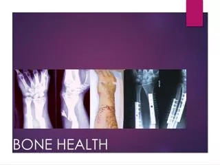
Measurement of Skeletal Status – 2011 Bone quality High-resolution MRI Micro-CT (from biopsy specimens) Hip structural analysis (bone geometry) Fracture rates Bone density • Dual energy x-ray absorptiometry (DXA) – 2D • Quantitative ultrasound (QUS) • Quantitative CT – 3D (including pQCT) • High-resolution pQCT (XtremeCT) • Peripheral vs. axial (central) measurements
DXA Terminology:Consider Different Regions of Skeleton • Central skeleton (axial skeleton plus hips and shoulders): • - Spine, ribs, pelvis, hips, shoulders • Peripheral skeleton (appendicular skeleton minus hips and shoulders): • - Extremities (arms and legs)
Definition of “osteoporosis” in children • No WHO definitions in children and teens • Concern for low bone mass • BMD Z-score by DXA < -2.0 SD • Slightly low if Z-score between -1.0 and -2.0 • “Diagnosis of osteoporosis in children and adolescents should NOT be made on the basis of BMD alone.” - Int’l Soc Clinical Densitometry 2007
Radial and Tibial Measurements Peripheral QCT Quantitative Ultrasound
XCT 3000 Radius Tibia Peripheral quantitative computed tomography of radius and tibia
Bone Turnover Cycle – hormonal balance enables appropriate activity of osteoblastsvs osteoclasts Bone Formation GH IGF-1 DHEA Androgens Bone Resorption Estrogen PTH Cortisol
What can we do as health care providers? • Rule out systemic disease, endocrinopathy bone loss • Amenorrhea in young woman be concerned! • Consider BMD measurement in at risk patients and ones with strong family history • Recall role of genetics in BMD determination • Encourage: • Regular exercise • Maintenance of normal weight • Good nutrition, with adequate calcium and vitamin D • Wean of glucocorticoids as primary disease allows
Diagnostic Work-Up Other: Ceruloplasmin, copper, IGF-I, DHEAS Bone age Urinary calcium/creatinine (spot/24 h) If amenorrhea: thyroid function, FSH, prolactin • Rule-out systemic disease • Consider insidious celiac disease • 25-hydroxyvitamin D • PTH • Calcium, phosphorus, magnesium
When should you order DXA scans? • Patients with multiple fractures • Pathologic (atraumatic fractures) • Diseases associated with skeletal deficiency states • Hypothalamic amenorrhea: after 6 months of amenorrhea • Be suspicious of low BMD if strong family history • Repeat scans only annually (except as part of research protocol)
US Office of Women’s Health Campaign: Best Bones Forever • www.bestbonesforever.gov • for girls • www.bestbonesforever.gov/parents for parents and partners
Thank you! Questions/Comments?