Download
1 / 92
920 likes | 1.2k Views
Aortic Emergencies. David Peterson February 3, 2005. Objectives. Define aortic dissection + AAA and demonstrate their clinical relevance Review pathophysiology and classification Discuss diagnostic modalities Discuss management options Series of questions and cases. PERSPECTIVE.

E N D
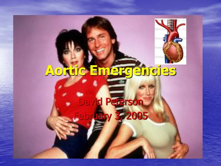
Aortic Emergencies David Peterson February 3, 2005
Objectives • Define aortic dissection + AAA and demonstrate their clinical relevance • Review pathophysiology and classification • Discuss diagnostic modalities • Discuss management options • Series of questions and cases
PERSPECTIVE • 1955 DeBakey outlined principles that remain basis for surgical treatment of aortic dissection • Medical treatment of aortic dissection was first advocated in the 1960s and is indicated for certain types of dissections
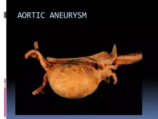
“longitudinal cleavage of the aortic media created by a dissecting column of blood” Rosen’s Emergency Medicine Aortic Dissection
(A) Normal blood flow in the aorta. (B) Dissection occurs when the inner lining of the aorta tears and the blood flow ‘dissects’ between the layers of the aortic wall
Aortic Dissection John Ritter Age 54 DEAD
Epidemiology • Underreported • Hypertension is the most common risk factor associated with aortic dissection and is seen in most patients • Incidence 5-10/ 1,000,000 and rising • Mortality 1-2%/hour (33% in 1st 24 hrs) • High rate of misdiagnosis ~28% • One study suggests EP’s suspect AD in <50% of cases • Sullivan et al. Am J Emerg Med 18: 46-50. 2000 • Variable presentation including MI • Lack of suspicion for AD is #1 cause of misdiagnosis
Epidemiology • History of cardiac surgery present 18% and bicuspid aortic valve in 14% of all patients with aortic dissection • more often in proximal dissections • Atherosclerosis rarely involved at site of dissection • Aortic dissection uncommon before age 40 except: • Congenital heart disease, Ehlers-Danlos or Marfan syndrome, giant cell arteritis and possibly pregnancy • ~ 44% of patients with Marfan syndrome develop aortic dissection and account for 6% of such cases
Anatomy and Physiology • Each contraction the heart swings side to side resulting in flexion of both ascending aorta and descending aorta • Descending aorta flexes just distal to left subclavian artery where mobile aorta is tethered • At 70 heartbeats per minute sequence occurs 37 million times a year causing repetitive stress on the aorta
Anatomy and Physiology • The aortic wall has three distinct layers: the intima, the media, and the adventitia • The media is comprised of elastic tissue and smooth muscle • MEDIA = Middle • INTIMA = Inside
Requires 3 basic features: Abnormal media Blood entry into media by intimal tear Pressure forces favoring propagation Pathophysiology
Pathophysiology • Arterial Obstruction is caused by 2 mechanisms • By compressing from the expanding hematoma • From occlusion of lumen by intimal flap • This results in peripheral complications: • Cerebral CVA, syncope • Spinal Neuro deficits • Cardiac MI, tamponade, AR • Respiratory Hemoptysis, pleural effusion • GI Hematemesis, dysphagia, mesenteric ischemia • Renal ARF, HTN • Limbs Extremity ischemia
Pathophysiology • Intimal tears occur in 96% of all AD cases • Felt to occur 2o to shearing forces and hemodynamic stresses • Propagation factors: • Degree of HTN • Slope of pulse wave (dP/dT) • Spontaneous cure = rupture back into true lumen (rare)
Aortic dissection bursts into the pericardium • Pericardial Tamponade
Hemopericardium and tamponade can occur with dissection into pericardial sac
The tear progresses down to and occludes the coronary artery precipitating an MI • Almost always an inferoposterior MI : RCA
Coronary artery involvement in ~1% presents as MI • 0.1-0.2% of MI’s are complicated by incorrect admin of lytics in • setting of AD
The tear can disrupt the aortic valve, producing acute valvular insufficiency, a common cause of acute CHF and increased risk of death AR
Clinical Presentation • Sudden, severe chest pain (~76-90%) • Migratory CP is highly specific (~71%) • Back pain (~53%), abd pain • Other Sx depending on site of involvement • Syncope (~9%) • Neuro Sx (~6-13%) • Mesenteric ischemia • Renal failure • Can be painless in up to 15% (chronic) Moore et al. Am J Card. 89:1235-1238 2002 Hals. Emerg Med Reports 2000
Risk Factors for AD • Hypertension (60-90%) • Age 50-70 yo • Male (3:1) • CTD’s (Marfans ~5%, Ehlers-Danlos) • Turners, Coarctation, Ebstein’s Anomaly • Congenital bi-/tricuspid AV • Family Hx or previous dissection • Cocaine, metamphetamine • Iatrogenic • Trauma Hals. Emerg Med Reports 2000
Classification • DeBakey: • Type I: involve ascending aorta, arch, and descending aorta • Type II: ascending aorta proximal to L subclavian artery • Type IIIa: descending aorta only; above diaphragm only • Type IIIb: descending aorta only; extension below diaphragm
Stanford • Type A: involvement of ascending aorta • Type B: no involvement of ascending aorta • ~62.5%of pts w/ AD have aType A • Involvement of ascending aorta is of prognostic and therapeutic importance
Diagnosing AD • Clinical suspicion above all • 3 clinical variables shown to be useful: • Aortic pain (immediate onset, tearing, ripping) • Mediastinal widening / aortic widening on CXR • Pulse or BP differentials • Likelihood of AD: • Ø of above variables 7% risk of AD • Pain or widening alone 31 + 39% risk of AD respectively • > 2 variables or isolated BP / pulse diff > 83% risk of AD • Von Kodolitsch et al. Arch Intern Med. 160: 2977-82. 2000 • Diagnostic modalities • ECG, CXR, CT, TEE, Angiogram, MRI
ECG findings in AD • ~85% will be abnormal • LVH • Non-specific ST-T wave changes • MI (RCA most common) • Bottom line: • not sensitive or specific • beware thrombolysis until AD excluded
CXR findings in AD • 80-90% will be abnormal • Most findings non-specific: • Mediastinal widening (~59-75%) • Calcium sign (pathognomonic) • Double density aorta • Obliteration of aortic knuckle • Loss of PA window • Tracheal deviation to right • Depressed left main stem bronchus • New left pleural effusion • Apical cap • Size disparity of ascending + descending aorta
TEE • Rosen: 98% sensitive, 77% specific • Moore et al: 88% sensitive • 1st test in Europe and Japan • Advantages: • Can differentiate Type A + B dissections • Rapid, can be done at bedside • No contrast or radiation • Can detect AR and pericardial effusion • Disadvantages • Availability, operator dependence • Limited info on distal aorta Moore et al. Am J Card. 89:1235-1238. 2002
TEE : Aortic Dissection Intra-operative TEE echo demonstrating high velocity jet striking aortic wall at point of intimal injury
CT scanning • Dynamic helical CT nearly 100% sensitive and specific (dye) (Moore et al: 93% sens) • Advantages: • Availability, can differentiate Type A + B • Able to identify sealed-off false lumens • Able to identify other pathology (eg PE) • Disadvantages: • Dye reactions (1/10,000 fatal) • No info on AV function or intimal tear location • No info about extension into other arteries
CT : Aortic Dissection CT of abdominal aorta show intimal flap (dark line)with true lumen anteriorly and false lumen posteriorly
CT : Aortic Dissection Stanford Type A Aortic Dissection
CT : Aortic Dissection Stanford Type A Aortic Dissection
Angiography • 81-87% sensitive, 96% specific • Previous gold standard • Advantages: • Anatomical delineation of aortic tree • Ability to demonstrate AR • Can differentiate Type A + B dissections • Disadvantages: • False –ves due to false lumen thrombosis • Invasive, time consuming, expensive
MRI • Near 100% sensitivity and specificity • No role in critical pts but good for serial follow-up • Advantages: • Excellent anatomical delineation, info on AR, intimal tear location, type and extent of AD • Disadvantages: • Time-consuming • Unable to monitor pt
Intravascular U/S • New technique – intravascular U/S probe • Currently evolving uses include • Identification of unstable plaques in CAD • Diagnostic and therapeutic use in AD • 3-D imaging of aorta + surrounding structures • Guidance of intra-vascular stent placement – less invasive procedure than classic surgery • Fenestration of intimal flap • Not available at most centers at this time Chavan et al. Circulation 96: 2124-2127. 1997
So what test do I order 1st? • Moore et al: CT is initial test of choice followed by TEE to clarify Dx or better delineate surrounding anatomy + AR • Moore et al. Am J Card. 89:1235-1238. 2002 • In Calgary, TEE is considered highly accurate and available, and should be considered a 1st line test • Peter Giannacarro
Can we predict outcomes? • Pulse deficit is independent predictor of 5-day mortality RR 2.73, 95% CI 1.7-4.4 • Stat sig trend of increasing mortality with increasing number of pulse deficits • Bosssone et al. Am J Card 89: 851-855. 2002 • Mortality predictors: • Age > 70 (OR 1.7, 95% CI 1.05-2.77) • Abrupt onset CP (OR 2.6, 95% CI 1.22-5.54) • Hypotension/shock/tamponade (OR 2.97, 95% CI 1.25-3.29) • ARF (OR 1.77; 95% CI 1.80-12.6) • Pulse deficit (OR 2.03; 95% CI 1.25-3.29) • Abnormal ECG (OR 1.77; 95% CI 1.06-2.95) • Rajendra et al. Circulation. 105: 200-206. 2002
Management • ABC’s and then 2 basic principles: • Control the slope of pulse wave (dP/dT) • 1st control your HR, then lower the pressure • Surgery if indicated • All Type A dissections need urgent OR • Controversies + New Ideas • Surgery for Type B dissections • Intravascular repair
Medical Tx • Control HR • IV BB’s – aim for HR 60-80 bpm • Esmolol 500 mcg/kg bolus, then titrate infusion 50-200 mcg/kg/min • Metoprolol 5 mg IVP q5min • Propranolol 1mg IVP q5min
Medical Tx • Control BP • aim for BP 100-120 mmHg sys or min BP req’d to maintain end-organ perfusion • Nitroprusside 0.5 mcg/kg/min – titrate up prn • Monotherapy • Labetalol 20 mg IVP, then 20-80 mg q5-10 min until in target HR, then 1-2 mg/hr
Surgical Tx • Indicated for: • All Type A dissections • Type B w/ complications: • Aortic rupture • Severe distal ischemia • Refractory HTN • Progressive dissection despite Tx • Intractable pain • Mortality for Type A repair is ~7-12% • Co-morbidities increase mortality • 5 yr survival is 77% Type A + 88% Type B