Download
1 / 20
201 likes | 270 Views
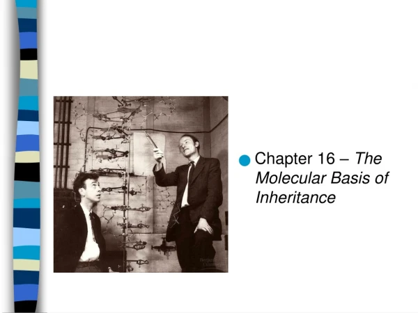
Chapter 16 – The Molecular Basis of Inheritance. Searching for Genetic Material. Mendel: modes of heredity in pea plants (1850s) Morgan: genes located on chromosomes (1910s)

E N D
Searching for Genetic Material • Mendel: modes of heredity in pea plants (1850s) • Morgan: genes located on chromosomes (1910s) • Frederick Griffith: bacterial work; identified the concept of transformation – a change in genotype and phenotype due to assimilation of an external substance (DNA) by a cell (1928) • Oswald Avery: transformation agent was DNA (1944) • Before Avery, many scientists believed that protein was the genetic material and not DNA. Can you think of a reason for this theory? • Protein is more complex (20 AAs as compared to 4 nucleotides) and a simple DNA strand couldn’t possibly give rise the complexity of humans.
Griffith’s Experiment • 2 strains of Streptococcus pneumoniae – 1 with a polysaccharide coat (smooth or S), one without (rough or R) • Conducted 4 experiments (fig. 16.2) • Injected mice with live S, the mice died, so encapsulated strain was pathogenic (disease causing) • Injected mice with live R, the mice stayed healthy, so the strain was non- pathogenic • Injected mice with heat-killed S, mice healthy, the polyscaaharide coat was found to not be the disease-causing agent because the coat was still intact in the heat-killed S bacteria • Heat killed S mixed with R and injected into mice, the mice died, the blood contained live S strain cells. The R strain “acquired” from S the ability to make a polysaccharide coat. These S cells were cultured and found to produce encapsulated daughter cells, concluded that newly acquired trait was inheritable. • The change in phenotype was due to an assimilation of external genetic material by a cell – transformation • Concluded that this could not be a protein because heat kills (denatures) proteins
Hershey and Chase • 1952 – discovered that DNA is the genetic material of a phage called T2 • T2 phage can infect E.coli, which is simply DNA enclosed by a protein coat, and could quickly reprogram the E.coli to produce new T2 phages. • Conducted experiments to figure out if the DNA or the protein was responsible for this reprogramming phenomenon • Phage protein was tagged with 35S, phage DNA was tagged with 32P • Allowed phage to infect separate samples of non-radioactive E. coli cells. • Centrifuged the samples- the heavier bacteria would fall to the bottom while the light virus would be on top • Measured radioactivity • Tubes with protein-labeled T2 had radioactivity in supernatant (liquid) • Tubes with DNA-labeled T2 had radioactivity in the pellet (bacteria) • Phage proteins remained outside the bacterial cells during infection, while phage DNA entered the cells • When cultured, bacterial cells with radioactive phage DNA released new phages with some 32P DNA • Concluded that DNA is the hereditary material of T2, not protein
DNA Structure • Edwin Chargaff (1947) – figured out ratio of nitrogen bases in a DNA molecule, approximate numbers of T=A and C=G, Chargaff’s rule • Rosalind Franklin (1950’s) – used X-ray crystallography to determine that DNA is in the form of a double helix
DNA Structure • Watson and Crick (1953) – proposed a model for DNA • Used x-ray crystallography data from Franklin as well a Chargaff’s rule to propose their model: • The structure is a double helix with a sugar-phosphate backbone and nitrogen bases in the center • Their model was not widely accepted until they could demonstrate how DNA could be replicated (i.e. its functionality) • In 1954, Watson and Crick published a second paper outlining how the process occurs – demonstrated that DNA replication is semiconservative • Data supported by Maurice Wilkins in 1956 • Watson, Crick, and Wilkins win the Nobel Prize in 1962 (Franklin not awarded prize – deceased, 1958)
DNA structure (and RNA) • DNA structure animation
DNA Replication - semiconservative • Proposed by Watson and Crick - strands are complementary; nucleotides line up on template according to base pair rules • Each new double helix will have one “old or original” strand and one “new” strand
DNA Replication – semiconservative • Meselson and Stahl (late 1950s) designed an experiment to test the theory of semiconservative DNA replication
DNA Replication Bubble • Origins of replication – a replication bubble • Begins at specific sites where parental strands separate with helicase enzyme and form bubble, stabilized with single-strand binding proteins • Each end of the bubble is a replication fork • Bubble expands laterally, DNA replication occurs in both directions • Eventually bubbles fuse, replication complete
DNA Replication Nucleotide Addition • New nucleotides are added to the free 3’ end of the growing strand (DNA formed in the 5’ to 3’ direction) • Utilizes a variety of polymerase enzymes to compete this addition • DNA is antiparallel, so how replicate both sides, simultaneously, in the same direction?
Leading and Lagging Strands • DNA Polymerase III adds new nucleotides to 3’ end • Leading strand – continuous replication towards the replication fork • Lagging strand – discontinuous replication away from the replication fork, replicated in chunks called Okazaki fragments • Animation overview
Lagging Strand • All replication begins with primase adding RNA nucleotides to DNA • DNA poly III adds nucleotides to the RNA primer sequence • DNA poly III releases when reach next RNA primer • DNA poly I replaces the RNA with DNA adding to 3’ end • DNA ligase forms bond between these two Okazaki fragments to complete DNA chain
Replication Overview • Overview
DNA Repair • Enzymes proofread DNA for errors • 1 in 10,000 initial errors, 1 in 1 billion end errors • Mismatch repair – occurs while being synthesized, uses DNA polymerase • Excision repair – corrects accidental changes to DNA, uses nuclease, polymerase, and ligase
DNA Shortening • On end of DNA is an RNA primer • Since DNA poly can only add to 3’ end, this is a problem • Because of this the ends get shorter • Telomeres – noncoding regions of DNA at end of chain, in humans repeating chains of TTAGGG • Protects genes from being eroded from successive rounds of replication • Telomerase catalyzes the length of telomeric DNA restoring the DNA length in germ cells; usually inactive in adult somatic cells