Download
1 / 34
390 likes | 777 Views
Primary Biliary Cirrhosis & Hemochromatosis . Aliyu Ndajiwo #605. Primary Biliary Cirrhosis. PBC. What is PBC?.

E N D
Primary Biliary Cirrhosis & Hemochromatosis Aliyu Ndajiwo #605
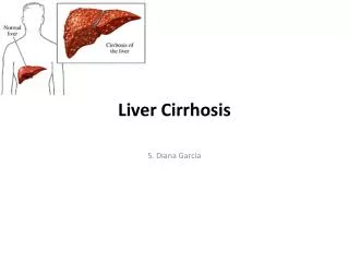
What is PBC? • Primary biliary cirrhosis, often abbreviated PBC, is an Autoimmune disease of the liver marked by the slow progressive destruction of the small bile ducts of the liver, with the interlobular ducts (Canals of Hering) affected early in the disease. • When these ducts are damaged, bile builds up in the liver (cholestasis) and over time damages the tissue. This can lead to scarring, cirrhosis.
Signs and Symptoms • Around half of all cases of primary biliary cirrhosis (PBC) have no symptoms until extensive liver damage has already occurred. This type of PBC is known as asymptomatic PBC Early-stage PBC: In people with symptoms, the most commonly reported symptoms of PBC are: • chronic fatigue (extreme tiredness) • Pruritis/itchy skin (ranging from mild to severe) • Dry eyes and mouth
Advanced PBC After PBC has caused extensive damage to the liver, there will be additional symptoms as a result of the liver not working as well as it should. These symptoms include: • jaundice • Edema • Ascites • Xanthoma, xanthelasmata • diarrhea – your stools may have a particularly unpleasant smell Physical examination findings depend on the stage of the disease. In the early stages, examination findings are normal. As the disease advances, the following signs may be noted: • Hepatomegaly (25%) • Hyperpigmentation (25%) • Splenomegaly (15%) • Jaundice (10%) • Xanthelasmata (10%): In late stages of the disease • Sicca syndrome (50-75%): Xerophthalmia (ie, dry eyes), xerostomia (ie, dry mouth)
Etiology The exact cause of primary biliary cirrhosis (PBC) is unknown, although it is thought a combination of genetic and environmental factors may play a part. Autoimmune condition • In cases of PBC, the immune system sends specialised cells, known as T-cells, to the site of the bile ducts. T-cells usually kill bacteria and viruses. • The T-cells start to damage the surface of the bile ducts, which gradually become extensively scarred and disrupt the usual flow of bile. As the amount of bile in the liver increases, further scarring of the liver (cirrhosis) occurs. • Over time, high levels of cirrhosis cause the liver to lose some, and eventually all, of its function. As the liver plays a vital role in filtering out impurities from your blood, a loss of liver function is potentially fatal.
Other autoimmune conditions Having one autoimmune condition such as PBC can make it more likely you will have other autoimmune conditions. For example, people with PBC may also have: • autoimmune thyroid disease • an underactive thyroid(hypothyroidism) • rheumatoid arthritis • scleroderma • autoimmunehepatitis • Sjogren's syndrome • Raynaud's phenomenon • coeliac disease
Increased risk There is some evidence to suggest that certain things increase your chances of developing PBC. These risk factors are explained below. Gender: PBC occurs mostly in women. Less than one in 10 cases of PBC occur in men. One theory for this is that the immune system is upset by the female sex hormone, oestrogen. Family history: Having a close relative with PBC means you are more likely to develop the condition yourself. For example, if you are female and your mother has or has had PBC, you are much more likely to develop PBC than someone who has no relatives with the condition. However, the risk is still low.. Possible triggers: Certain things may trigger PBC in people with a genetic tendency. These include: • infections, such as a urinary tract infection (UTI) • hormone replacement therapy (HRT) – a treatment used to replace the female hormones that a woman's body no longer produces because of the menopause • using nail varnish or cosmetics • smoking cigarettes or having a past history of heavy smoking • living close to where toxic wastes are dumped However, none of these have been proven as definite triggers of PBC. Most people who are exposed to them will not develop the condition. At present, there is nothing to suggest you should reduce your exposure to these triggers to lower your risk of developing PBC.
Complication As liver damage progresses, people with primary biliary cirrhosis may develop a number of serious problems, including: • Increased pressure in the portal vein (portal hypertension). Blood from your intestine, spleen and pancreas enters your liver through a large blood vessel called the portal vein. When scar tissue blocks normal circulation through your liver, blood backs up, much like water behind a dam, leading to increased pressure within the vein. And because blood doesn't flow normally through your liver, hormones, drugs and other toxins aren't filtered properly from your bloodstream. • Enlarged veins (varices). When circulation through the portal vein is slowed or blocked, blood may back up into other veins — mainly those in your stomach and esophagus. The blood vessels are thin walled, and increased pressure in your veins can cause bleeding in your upper stomach or esophagus. This bleeding is a life-threatening emergency that requires immediate medical care. • Cirrhosis • Liver cancer. • Weak bones (osteoporosis). • Vitamin deficiencies. • Memory problems. • An increased risk of other disease. In addition to bile duct and liver damage, people with primary biliary cirrhosis are likely to have other metabolic or immune system disorders, including thyroid problems, limited scleroderma (CREST syndrome) and rheumatoid arthritis.
Diagnosis In many cases, a suspected diagnosis of primary biliary cirrhosis (PBC) is made when carrying out a blood test for another, unrelated condition. Blood tests • A diagnosis of PBC can usually be confirmed by checking your blood for a substance called anti-mitochondrial antibodies (AMAs). In PBC, AMAs are produced by the immune system (the body's natural defence against infection and illness). They are present in 95% of people with PBC. • You may also have a high level of bilirubin in your blood. Liver biopsy • You may also be referred for a liver biopsy. This involves a small sample of liver tissue being taken so it can be studied under a microscope. • A liver biopsy is not always necessary to diagnose PBC, which is usually confirmed by the presence of AMAs. However, it can be used to assess the extent of damage to your liver.
Staging: • Stage 1 (portal stage of Ludwig): Portal inflammation, bile duct abnormalities, or both are present • Stage 2 (periportal stage): Periportal fibrosis is present, with or without periportal inflammation or prominent enlargement of the portal tracts with seemingly intact, newly formed limiting plates • Stage 3 (septal stage): Septal fibrosis with active inflammatory, passive paucicellular septa, or both are present • Stage 4 (cirrhosis): Nodules with various degrees of inflammation are present
Figure 1: Histological staging in a representative case of a patient with primary biliary. Stage 1 reveals duct-centred inflammation showing chronic nonsuppurative destructive cholangitis (black arrows). A tiny granuloma is also seen (grey arrow). Stage 2 shows portal enlargement (arrows) with bile ductular reaction and inflammatory cell infiltration. Stage 3 is characterized by fibrous scaring bridging portal tracts with occasional foci of bile duct loss (no bile duct identified around an artery indicated by arrow). Stage 4 shows cirrhotic transformation.
Treatment and Drugs Because no cure exists for primary biliary cirrhosis, treatment focuses on slowing the progress of the disease, relieving symptoms and preventing complications. Self-help • Quit smoking (if you smoke) • Avoid obesity by maintaining a healthy weight • Do not drink more than the recommended daily limits of alcohol (3-4 units a day for men, and 2-3 units a day for women) Treating the disease: • Ursodeoxycholic acid (UDCA) - Also known as ursodiol (Actigall, Urso), UDCA is a bile acid that helps move bile through your liver. Although UDCA doesn't cure primary biliary cirrhosis, it may prolong life if started early in the disease. It’s less likely to help people with advanced liver damage. Side effects of UDCA may include weight gain, hair loss and diarrhea. • Liver transplant. When treatments no longer control primary biliary cirrhosis and the liver begins to fail, a liver transplant may help prolong life. Primary biliary cirrhosis often recurs in the transplanted liver, but it may take several years to develop
Treating the symptoms Your doctor may recommend treatments to control the signs and symptoms of primary biliary cirrhosis to make you more comfortable. Treatments may help control symptoms such as: • Fatigue. Treatment for fatigue involves trying to determine what may contribute to your symptoms. One medication that has shown promise in studies is Modafinil (Provigil). More research is needed to determine its role in primary biliary cirrhosis. • Itching. One option for controlling itching is cholestyramine (Locholest, Prevalite), which is a powder that must be mixed with food or liquids. Though cholestyramine works for most people, the taste is unpleasant. Another option is rifampin (Rifadin), which is taken in pill form. Rifampin doesn't work for everyone. Itching that can't be controlled may be treated with a liver transplant. Preventing complications • Increased pressure in the portal vein (portal hypertension). Your doctor is likely to screen for portal hypertension and enlarged veins when you're first diagnosed and every few years thereafter. If you're diagnosed with portal hypertension or bleeding, treatment may involve medications or surgery. • Weak bones (osteoporosis). • Vitamin deficiencies.
Epidemiology • The female:male ratio is at least 9:1. • In some areas of the US and UK the prevalence is estimated to be as high as 1 in 4000. This is much more common than in South America or Africa, which may be due to better recognition in the US and UK. • First-degree relatives may have as much as a 500 times increase in prevalence, but there is debate if this risk is greater in the same generation relatives or the one that follows.
Prognosis • The serum bilirubin level is an indicator of the prognosis of primary biliary cirrhosis, with levels of 2–6 mg/dL having a mean survival rate of 4.1 years, 6–10 mg/dL having 2.1 years and those above 10 mg/dL having a mean survival time of 1.4 years. • After liver transplant, the recurrence rate may be as high as 18% at 5 years, and up to 30% at 10 years. There is no consensus on risk factors for recurrence of the disease. • Patients with primary biliary cirrhosis have an increased risk of hepatocellular carcinoma.
References • http://www.mayoclinic.org/diseases-conditions/primary-biliary-cirrhosis/basics/definition/con-20029377 • http://www.nhs.uk/Conditions/Primary-biliary-cirrhosis/Pages/Introduction.aspx • http://en.wikipedia.org/wiki/Primary_biliary_cirrhosis
Hemochromatosis (Iron Overload)
Background Hemochromatosisis the abnormal accumulation of iron in parenchymal organs, leading to organ toxicity. This is the most common inherited liver disease in white persons and the most common autosomal recessive genetic disorder. • Normal reference ranges are: • Serum Iron (SI): • Men: 65 to 176 μg/dL • Women: 50 to 170 μg/dL • Newborns: 100 to 250 μg/dL • Children: 50 to 120 μg/dL • TIBC: 240–450 μg/dL • Transferring Saturation:20–50% *μg/dL = microgram per deciliter. • Excess iron is hazardous, because it produces free radical formation. The presence of free iron in biologic systems can lead to the rapid formation of damaging reactive oxygen metabolites, such as the hydroxyl radical and the superoxide radical. These can produce DNA cleavage, impaired protein synthesis, and impairment of cell integrity and cell proliferation, leading to cell injury and fibrosis.
Other types of hemochromatosis • Juvenile hemochromatosis: This causes the same problems in young people that hereditary hemochromatosis causes in adults. But iron accumulation begins much earlier, and symptoms usually appear between the ages of 15 and 30. This disorder is caused by a mutation in the HJV gene. • Neonatal hemochromatosis: In this severe disorder, iron builds up rapidly in the liver of the developing fetus. It is thought to be an autoimmune disease, in which the body attacks itself. • Secondary hemochromatosis: is caused by disorders of erythropoiesis and treatment of the diseases with blood transfusions. After damage of transfused erythrocytes by macrophages, iron freed from heme is accumulated in the body (liver, heart, skin). • Secondary hemochromatosis is mainly induced by diseases of erythropoiesis, including: • Thalassemia • Sickle cell anemia • X-linked sideroblastic anemia • Pyruvate kinase deficiency • Hereditary spherocytosis • Congenital dyserythropoietic anemia (CDA).
Signs and Symptoms • Patients with hereditary hemochromatosis may be asymptomatic (75%) or may present with general and organ-related signs and symptoms. • Early symptoms include: • Severe fatigue (74%), • Impotence (45%), • Arthralgia (44%). • The most common signs at the time of presentation are: • Hepatomegaly (13%) • Skin pigmentation (bronze) • Arthritis. When symptoms are associated with hemochromatosis, these usually begin in men in their late 20’s to early 30’s. In women, symptoms usually start about 10-15 years after they stop having a period due to menopause, birth control pills or hysterectomy
Etiology • Hereditary hemochromatosis is caused by a mutation in a gene that controls the amount of iron your body absorbs from the food you eat. The mutations that cause hereditary hemochromatosis are passed from parents to children. Gene mutations that cause hemochromatosis • The gene that is mutated most often in people with hereditary hemochromatosis is called HFE. You inherit one HFE gene from each of your parents. • The HFE gene has two common mutations, C282Y and H63D. One of these mutations is found in about 85 percent of people who have hereditary hemochromatosis. Genetic testing can reveal whether you have these mutations in your HFE gene. • If you inherit 2 abnormal genes, you may develop hemochromatosis. About 70 percent of people who inherit two genes develop evidence of iron overload of hemochromatosis. You can also pass the mutation on to your children. • If you inherit 1 abnormal gene, you won't develop hemochromatosis. You are considered a gene mutation carrier and can pass the mutation on to your children. They would not develop disease unless they also inherit another abnormal gene from another parent.
How hemochromatosis affects your organs Iron plays an essential role in several body functions, including helping in the formation of blood. A peptide hormone called hepcidin, secreted by the liver, plays a key role in the body's use of iron. It controls how much iron is absorbed by the intestines, how iron is used in various body processes and how it's stored in various organs. In hemochromatosis, the normal role of hepcidin is disrupted and your body absorbs more iron that it needs. This excess iron is stored in the tissues of major organs, especially your liver. Too much iron is toxic to your body, and over a period of years, the stored iron can severely damage many organs, leading to organ failure and chronic diseases such as cirrhosis, diabetes and heart failure. Though many people have faulty genes that cause hemochromatosis, only about 10 percent of them have iron overload to the degree that causes tissue and organ damage.
Risk Factors Factors that increase your risk of hereditary hemochromatosis include: • Having 2 copies of a mutated HFE gene. This is the greatest risk factor for hereditary hemochromatosis. • Family history. If you have a first-degree relative — a parent or sibling — with hemochromatosis, you're more likely to develop the disease. If you have a family history of alcoholism, heart attacks, diabetes, liver disease, arthritis or impotence, your risk of hemochromatosis is greater. • Ethnicity. People of Northern European descent are more prone to hereditary hemochromatosis than are people of other ethnic backgrounds. Hemochromatosis is less common in African-Americans, Hispanics and Asian-Americans. • Being a man. Men are more likely to develop signs and symptoms of hemochromatosis at an earlier age. Because women lose iron through menstruation and pregnancy, they tend to store less of the mineral than men do. After menopause or a hysterectomy, the risk for women increases.
Tests & Diagnosis Clinical manifestations of hemochromatosis include the following: • Liver disease (hepatomegaly (13%); cirrhosis (13%) usually late in the disease) • Skin bronzing or hyperpigmentation (70%) • Diabetes mellitus (48%) • Arthropathy • Amenorrhea, impotence, hypogonadism • Cardiomyopathy • Osteopenia and osteoporosis • Hair loss • Koilonychia (spoon nails) Lab tests The diagnosis of hemochromatosis is based on clinical features of the disease. Most patients are asymptomatic and are diagnosed when elevated serum iron levels are noted on a routine chemistry screening panel or when screening is performed because a relative is diagnosed with hemochromatosis. Laboratory studies used in evaluating suspected hemochromatosis include the following: • Genetic testing: Examination of HFE mutations (C282Y, H63D) is important for diagnosis of hemochromatosis • Transferrin saturation levels • Serum ferritin studies • Hepatic iron concentration Supraventricular arrhythmias are often revealed on ECGs.
Imaging studies Chest radiography and echocardiography may be helpful in the evaluation of patients with hemochromatosis and cardiac disease. CT scanning is neither sensitive nor specific for the detection of mild hepatic iron overload. MRI may be more sensitive than CT scanning, but this modality has not been validated as a diagnostic test to help confirm hemochromatosis. Hepatic iron quantification with MRI might be helpful. Procedures The following are procedures that may be used to assess patients with hemochromatosis: • Diagnostic endoscopy • Skin biopsy • Liver biopsy, with biochemical determination of hepatic iron concentration and calculation of the hepatic iron index and histologic evaluation with iron staining (Perls Prussian blue)
Management Clinical suspicion and early diagnosis are essential in hemochromatosis. The goal of therapy in patients with iron overload disorders is to remove the iron before it can produce irreversible parenchymal damage. Phlebotomy Once diagnosed, hemochromatosis is treated by phlebotomy to rid the body of excess iron and to maintain normal iron stores. Phlebotomy remains the sole recommended treatment for hereditary hemochromatosis and should be undertaken in a case-specific manner. Chelation therapy In patients with hemochromatosis and heart disease, anemia, or poor venous access, treatment with iron chelation agents is recommended and includes the following agents: • Deferoxamine - It binds with iron in the bloodstream and enhances its elimination via urine and faeces. Two newer iron chelating drugs that are licensed for use in patients receiving regular blood transfusions to treat thalassaemia (and, thus, who develop iron overload as a result) are : • Deferasirox • Deferiprone
Surgery Surgical procedures are used to treat 2 important complications of hemochromatosis: end-stage liver disease and severe arthropathy. Orthotopic liver transplantation is the only therapeutic option when end-stage liver disease progresses despite iron-reduction therapy. This intervention is also indicated in cases with the development of hepatocellular carcinoma. Surgical arthroplasty is considered if joint destruction becomes severe despite medical therapy.
Epidemiology • Prevalence of hereditary hemochromatosis in the United States is 1 case in 200-500 individuals. Most are of northern European origin • Hemochromatosis is less common among patients of African descent. • Marked racial disparity in the distribution of the C282Y mutation has been noted. Prevalence of hemochromatosis is 6 times higher in white persons than in black persons. In the Irish, the frequency of the C282Y mutation was 10%, whereas in Australian aboriginal, African, and Asian populations, the mutation has not been found. • Men are affected with hemochromatosis nearly 2-3 times as often as women • Hemochromatosis usually becomes apparent after age 40 years in men (median age, 51 y)and after age 50 years in women (median age, 66 y). In women, onset of hereditary hemochromatosis begins later because menstruation causes physiologic blood loss, which increases iron removal.
Prognosis The most important prognostic factor at the time of diagnosis is the presence or absence of hepatic fibrosis or cirrhosis. Patients without significant hepatic fibrosis may be expected to have a normal life expectancy with phlebotomy therapy. Adequate phlebotomy treatment is the major determinant of survival, and it markedly improves prognosis. Early diagnosis and therapeutic phlebotomy to maintain low normal body stores is crucial and can prevent all known complications of hemochromatosis. If untreated, hemochromatosis may lead to death from • Cirrhosis • Diabetes • Malignant hepatoma (Hepatocellualr carcinoma) • Or cardiac disease
References • http://www.mayoclinic.org/diseases-conditions/hemochromatosis/basics/definition/con-20023606 • http://en.wikipedia.org/wiki/Iron_overload • http://www.hemochromatosis.org/ • http://emedicine.medscape.com/article/177216-overview