Download

1 / 37
460 likes | 1.16k Views
Learn about hemoptysis, the expectoration of blood, its various causes from vascular to parenchymal disorders, and the importance of differential diagnosis. Understand the evaluation process to identify potential life-threatening conditions and receive appropriate treatment.

E N D
HEMOPTYSIS Suhail Allaqaband Sinai Samaritan Medical Center Milwaukee, WI
HEMOPTYSIS • Hemoptysis, or the expectoration of blood, can range from blood-streaking of sputum to the presence of gross blood in the absence of any accompanying sputum • The term massive hemoptysis is reserved for bleeding that is potentially life-threatening • It has been defined by a number of different criteria, often ranging from more than 100 to more than 600 ml of blood over a 24 hour period
VASCULAR ORIGIN OF HEMOPTYSIS • Blood traversing the lungs can arrive from • pulmonary arteries, or • bronchial arteries • Virtually the entire cardiac output courses through the low-pressure pulmonary arteries and arterioles en route to being oxygenated in the pulmonary capillary bed • In contrast, the bronchial arteries are under much higher systemic pressure but carry only a small portion of the cardiac output
VASCULAR ORIGIN OF HEMOPTYSIS • Despite the quantitatively smaller contribution of the bronchial circulation to pulmonary blood flow, the bronchial arteries are generally a more important source of hemoptysis than the pulmonary circulation • In addition to being perfused at a higher pressure, they also supply blood to the airways and to lesions within the airways
DIFFERENTIAL DIAGNOSIS OF HEMOPTYSIS • Before assuming a lower respiratory source of the bleeding, it is important to consider whether the blood may be coming from a non-pulmonary source, such as the upper airway or the gastrointestinal tract • Alkaline pH, foaminess, or the presence of pus may sometimes suggest the lungs as the primary source of bleeding rather than the stomach.
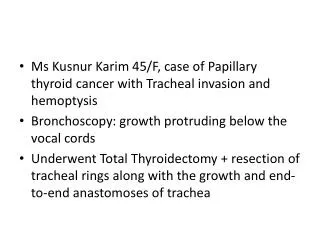
Airways diseases • The most common source of hemoptysis is airways disease • Inflammatory diseases, such as bronchitis or bronchiectasis • Neoplasms, including primary bronchogenic carcinoma, endobronchial metastatic carcinoma or bronchial carcinoid • In patients with AIDS, Kaposi's sarcoma involving the airways and/or the pulmonary parenchyma • Foreign body & Airway trauma • Fistula between a vessel and the tracheobronchial tree • fistulas between the aorta and the airway are associated with aneurysms of the thoracic aorta and are fatal if not diagnosed and surgically treated • Tracheo-innominate fistulas are a rare but potentially life-threatening complication of tracheostomy
Pulmonary parenchymal diseases • Infection, especially tuberculosis, pneumonia, aspergilloma, and lung abscess • Hemoptysis, which can be life-threatening, complicates the course of 50 to 85 percent of patients with an aspergilloma • Tuberculosis can cause massive hemoptysis through multiple mechanisms • Active cavitary or noncavitary lung disease can cause small or large amounts of bleeding • Active disease can cause sudden rupture of a Rasmussen's aneurysm (aneurysm of the pulmonary artery that slowly expands into an adjacent cavity because of inflammatory erosion of the external vessel wall until it bursts)
Pulmonary parenchymal diseases • Inflammatory or immune disorders • Goodpasture's syndrome, idiopathic pulmonary hemosiderosis, lupus pneumonitis, and Wegener's granulomatosis • Coagulopathy • thrombocytopenia or use of anticoagulants • Iatrogenic, especially due to either percutaneous or transbronchial lung biopsy • Hemoptysis, which is usually minor and transient, occurs in five to 10 percent of percutaneous lung biopsies, but massive hemorrhage and death have also been reported
Miscellaneous causes of pulmonary parenchymal hemorrhage • Cocaine-induced pulmonary hemorrhage • Hemoptysis has been described in six percent of habitual smokers of free-base cocaine ("crack") and has been associated with diffuse alveolar hemorrhage • Catamenial hemoptysis • hemoptysis that is recurrent and coincident with menses. The cause is intrathoracic endometriosis, usually involving the pulmonary parenchyma but occasionally affecting the airways
Pulmonary vascular disorders • Pulmonary embolism • Pulmonary AV malformation, either with or without underlying Osler-Weber-Rendu syndrome • Elevated pulmonary capillary pressure • mitral stenosis • significant left ventricular failure • Congenital heart disease • severe pulmonary hypertension • Iatrogenic • pulmonary artery perforation from a Swan-Ganz catheter
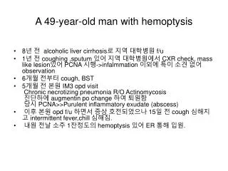
Cryptogenic • Depending upon the study, up to 30 percent of patients with hemoptysis have no cause identified even after careful evaluation • In a series of 67 patients with cryptogenic hemoptysis, the prognosis was generally good, and most patients had resolution of bleeding within six months of evaluation Adelman, M, et al. Cryptogenic hemoptysis. Clinical features, bronchoscopic findings, and natural history in 67 patients. Ann Intern Med 1985; 102:829
EVALUATION OF HEMOPTYSIS • The evaluation should begin with the initial history and physical examination supplemented by chest radiograph • Important features of the history include age, smoking history, duration of hemoptysis, and association with symptoms of acute bronchitis or an acute exacerbation of chronic bronchitis
Important historical points to address • Is there a history of prior lung, cardiac, or renal disease? • Is there a history of cigarette smoking? • Has the patient had prior hemoptysis, other pulmonary symptoms, or infectious symptoms? • Is there a family history of hemoptysis or brain aneurysms (suggesting hereditary hemorrhagic telangiectasia)? • Is there a history of skin rash? • What is the patient's travel history? • Does the patient have a history of asbestos exposure? • Is there a history of bleeding disorders or use of aspirin, nonsteroidal anti-inflammatory drugs, or anticoagulants? • Is there a history of upper airway or upper gastrointestinal complaints or diseases?
Physical examination • The presence of many telangiectasias suggests HHT • A skin rash may be suggestive of vasculitis • Splinter hemorrhages suggest endocarditis or vasculitis • Clubbing is nonspecific, since it can occur in many chronic lung diseases • Pulmonary hypertension may be suggested by an augmented P2, murmurs of tricuspid regurgitation or pulmonic insufficiency, or a right ventricular lift • Cardiac murmurs also raise the question of congenital heart disease, endocarditis with septic emboli, or, when a diastolic rumble or opening snap is present, mitral stenosis • The legs should be examined carefully for possible deep venous thrombi
EVALUATION OF HEMOPTYSIS • No immediate further work-up is indicated if the clinical picture is not suggestive of carcinoma • negative chest radiograph, • age less than 40 years, • no smoking history, and • hemoptysis less than 1 week duration but • Is suggestive of acute bronchitis (blood streaking superimposed upon purulent sputum) • Such a patient should be treated for bronchitis and observed for recurrence of hemoptysis following improvement in purulent sputum production
LABORATORY EVALUATION • Additional studies which may be useful depending upon the particular clinical situation include • hematocrit, urinalysis, blood urea nitrogen and plasma creatinine concentration, a coagulation profile, and collection of sputum for cytologic and microbiologic studies • Serologic tests for Wegener's granulomatosis, SLE, or Goodpasture's syndrome may be very helpful if positive • An echocardiogram may detect endocarditis, mitral stenosis, congenital heart disease, or pulmonary hypertension • A transesophageal echocardiogram may identify a thoracic aortic aneurysm as the cause of hemoptysis
EVALUATION OF HEMOPTYSIS • Further evaluation is indicated if the patient has risk factors for carcinoma or if the hemoptysis does not occur in the setting of acute bronchitis • Bronchoscopy is the preferred next procedure in those patients with risk factors for tumor or chronic bronchitis • On the other hand, HRCT is the preferred next procedure in patients at lower risk for tumor or chronic bronchitis but with a history or radiograph suggestive of bronchiectasis or an arteriovenous malformation.
DIAGNOSTIC PROCEDURES • Fiberoptic bronchoscopy • often considered in patients with hemoptysis and a normal or nonlocalizing CXR to rule out endobronchial malignancy • performed early in the evaluation, while the patient is actively bleeding, provides the highest yield for localizing the bleeding site • Risk factors predicting those individuals most likely to have tumor found on bronchoscopy include • Male sex • Older age, greater than 50 years • Smoking history greater than 40 pack years. • Duration of hemoptysis greater than one week
Arteriography • If the patient continues to bleed and the source is still unknown, then arteriography should next be performed, since it may be useful for therapy as well as diagnosis • Since the majority of massive bleeds arise from the bronchial circulation, bronchial arteriography has a higher yield than arteriography of the pulmonary or systemic arterial beds • When the pulmonary arterial circulation is the source, the most common underlying conditions are pulmonary AVM’s, Rasmussen's aneurysms or iatrogenic pulmonary artery tears
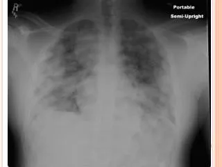
Computed tomography of the chest • Use of early chest CT has been advocated to help localize the bleeding site and diagnose the cause of hemoptysis • The advantage of CT is that it may suggest one of several diagnoses, such as bronchiectasis, lung abscess, and mass lesions, including cancer, mycetomas, and AVM’S • It may also help in the acute setting to guide arteriography or bronchoscopy to the regions of highest yield • The disadvantage of chest CT is that it may require temporary movement of an unstable patient away from intensive care
Multidetector Helical CT Aortogram
Fiberoptic bronchoscopy versus HRCT • Fiberoptic bronchoscopy and HRCT are, in many ways, complementary studies, each with specific advantages in certain clinical situations • In one study of 91 patients with hemoptysis, HRCT demonstrated all tumors seen by bronchoscopy as well as several which were beyond bronchoscopic range. On the other hand, HRCT could not detect bronchitis or subtle mucosal abnormalities which could be seen by bronchoscopy • In another report of 57 patients, HRCT was particularly useful in diagnosing bronchiectasis and aspergillomas, while bronchoscopy was diagnostic of bronchitis and mucosal lesions such as Kaposi's sarcoma
ACUTE MANAGEMENT • Initial priorities are insuring adequate airway protection, ventilation, and cardiovascular function • Patients with poor gas exchange, rapid ongoing hemoptysis, hemodynamic instability, or severe shortness of breath should be orally intubated with a large bore endotracheal tube (size 8.0 or greater) • Coagulation disorders should be rapidly reversed.
Protection of the nonbleeding lung • If the location or side of bleeding is known, placing the bleeding lung in a dependent position may prevent blood spillage into the nonbleeding lung • An alternative strategy involves placement of a typical, single lumen endotracheal tube into either the right or left mainstem bronchus • The approach of selective intubation is less practical when the right lung is bleeding, because selective intubation of the left mainstem bronchus may be quite difficult • A third alternative is the placement of a double lumen endotracheal tube specially designed for selective intubation of the right or left mainstem bronchi
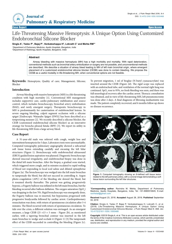
Use of bronchoscopy in acute management • A bronchoscopic option for protecting the non-bleeding lung is balloon tamponade of the bleeding site, involving placement of a Fogarty balloon catheter in the segmental or subsegmental bronchus leading to the bleeding site • The balloon is left inflated for 24 to 48 hours, and the patient is then observed for rebleeding with the balloon deflated for several hours • There is a potential risk of ischemic mucosal injury and postobstructive pneumonia, but these complications have not been reported
Use of bronchoscopy in acute management • Bronchoscopic techniques used to slow or stop bleeding have included lavage with iced saline and application of topical epinephrine (1:20,000), vasopressin, thrombin, or a fibrinogen-thrombin combination • None of these methods has been tested in controlled trials • If bronchoscopy visualizes a localized bleeding mucosal lesion, laser therapy or electrocautery may be considered, if available
Surgery • Patients with lateralized, uncontrollable bleeding should be assessed early for possible surgery • Relative contraindications to surgery include severe underlying pulmonary disease, active TB, diffuse underlying lung disease (cystic fibrosis, multiple AVMs, multifocal bronchiectasis), and diffuse alveolar hemorrhage • Morbidity and mortality are significantly greater with emergent surgery for persistent massive bleeding compared with elective surgery • In most series of emergent therapy, surgical mortality for treatment of massive hemoptysis is approximately 20 %
Arteriographic embolization • The other option for the patient who continues to bleed is arteriographic embolization, either as "semi-definitive" treatment or as a bridge to elective surgery • In the hands of experienced angiographers, embolization successfully stops bleeding more than 85 percent of the time • Unfortunately, embolization is only "semi-definitive," because rebleeding occurs in 10 to 20 percent of patients over the next 6 to 12 months • Late rebleeding may be due to incomplete embolization, revascularization, or recanalization.
RECOMMENDATIONS • First, stabilize the patient and then perform early bronchoscopy along with other appropriate diagnostic studies • If the patient continues to bleed aggressively, arteriography is most reasonable for localization and therapy • If bleeding persists despite embolization or if the patient is too ill to go to angiography, then blockade therapy or a double lumen tube should be considered • While surgery remains the only truly definitive therapy, it should not be used in the acute emergent setting unless it cannot be avoided