Download

1 / 142
1.41k likes | 1.5k Views
Medical school pathology lectures, year review of term 4 - Kidney, Urogenital system and tropical infections.

E N D
Pathology Review-Term4 Tip for Success in life….! "Powered by intellect, Driven by Values..!” Life motto of Infosys founder and Chairman, Narayana Murthy. INDIA 1
T4W1: Week overview: 2013 Term 4 CPC 1 Title: MSK System: Rheumatology Aim: To train students in: History taking + clinical examination of patient with joint pain ; pathology of physiology of rheumatological diseases; process of care + population health especially in rural and remote areas Learning 1. Demonstrate competency in history taking & clinical examination Outcomes: of patients Students will be presenting with joint pain able 2. Describe the Pathophysiology of to • Rheumatoid arthritis (RA) • Sero-negative arthritis • Osteoarthritis (OA), • Gout 3 Describe differential diagnoses for patients presenting with joint pains. 4 Formulate a first line management plan for patients presenting with joint pains, demonstrating a knowledge of indications and side effects of commonly used medications prescribed for treatment of joint pain. T4W1: Week overview: 2013 Term 4 CPC 1 Title: MSK System: Rheumatology Aim: To train students in: History taking + clinical examination of patient with joint pain ; pathology of physiology of rheumatological diseases; process of care + population health especially in rural and remote areas Learning 1. Demonstrate competency in history taking & clinical examination Outcomes: of patients Students will be presenting with joint pain able 2. Describe the Pathophysiology of to • Rheumatoid arthritis (RA) • Sero-negative arthritis • Osteoarthritis (OA), • Gout 3 Describe differential diagnoses for patients presenting with joint pains. 4 Formulate a first line management plan for patients presenting with joint pains, demonstrating a knowledge of indications and side effects of commonly used medications prescribed for treatment of joint pain.
CPC 4.1 – MSK-Rheumatology – Scenario 1: Rheumatoid A Ms F.M. 19 year old student Gouty Arthritis Mr J.W. 45 year old foot, 1st metatarsal – Scenario 2: Osteoarthritis Mrs N.M 69y retired Sports teacher. – Scenario 3: • Notes to Tutors: – Discuss DD - variety of clinical scenarios. – Remember/revise serious causes of acute joint pain esp. septic arthritis, rheumatic fever (Jones criteria). – DD to include fibromyalgia, polymyalgia rheumatica, SLE etc. • Investigations: – FBC, RFT, ESR/CRP, ALP, Auto Ab.P, RF & Anti-CCP, HLA B27, 3 CPC 4.1 – MSK-Rheumatology – Scenario 1: Rheumatoid A Ms F.M. 19 year old student Gouty Arthritis Mr J.W. 45 year old foot, 1st metatarsal – Scenario 2: Osteoarthritis Mrs N.M 69y retired Sports teacher. – Scenario 3: • Notes to Tutors: – Discuss DD - variety of clinical scenarios. – Remember/revise serious causes of acute joint pain esp. septic arthritis, rheumatic fever (Jones criteria). – DD to include fibromyalgia, polymyalgia rheumatica, SLE etc. • Investigations: – FBC, RFT, ESR/CRP, ALP, Auto Ab.P, RF & Anti-CCP, HLA B27, 3
Differentiating Features: Rheumatoid Arthritis: • Young, small joints • Autoimmune. • Synovial Inflammation • synovium Cartilage Osteoarthritis: • Old age, Large joints • Degenerative. • Cartilage degeneration. • Cartilage Synovium 5 Differentiating Features: Rheumatoid Arthritis: • Young, small joints • Autoimmune. • Synovial Inflammation • synovium Cartilage Osteoarthritis: • Old age, Large joints • Degenerative. • Cartilage degeneration. • Cartilage Synovium 5
Degenerative - Inflammatory • • • • • • • Both sexes equal. Pain through the day No morning stiffness. Stiffness, less pain. Bony swelling. No soft tissue swelling Uni/Bilateral, Asymmetrical. • • • • • Females more. Morning stiffness >1h. Less with movement. Pain & redness Inflammation & swelling of soft tissue. • Late bone swelling. • Bilateral, Symmetrical. 6 Degenerative - Inflammatory • • • • • • • Both sexes equal. Pain through the day No morning stiffness. Stiffness, less pain. Bony swelling. No soft tissue swelling Uni/Bilateral, Asymmetrical. • • • • • Females more. Morning stiffness >1h. Less with movement. Pain & redness Inflammation & swelling of soft tissue. • Late bone swelling. • Bilateral, Symmetrical. 6
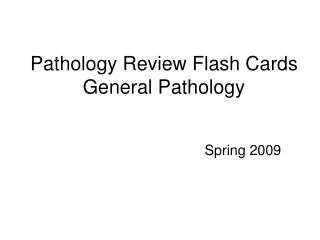
Normal -- Femur Head -- OA Normal Osteoarthritis 8 Normal -- Femur Head -- OA Normal Osteoarthritis 8
Osteoarthritis: Ankylosis • varus deformity of the knee and collapse of the joint space with destruction of the medial cartilage and the subchondral cortex (open arrowheads). 9 Osteoarthritis: Ankylosis • varus deformity of the knee and collapse of the joint space with destruction of the medial cartilage and the subchondral cortex (open arrowheads). 9
. RA - Pathophysiology Autoimmune TH1, TH17 & B cells MMPs, TNF, PGE2 Synovial Inflam & Proliferation with Papillary projections Chronic Inflam. Lymphocytes & Lymphoid follicles 11
. Gout: • 1%, Males common, • High serum uric acid + monosodium urate crystals in & around joints. – Primary 90% - congenital – Secondary 10% (malignancies, renal disesase, high protein diet) – Acute / Chronic. Tophus • Large deposits – Tophi Acute Chronic 13
. Degenerative Disc Disease (DDD) • Ageing / trauma • Low back pain/deformity. • Complication: Common Disc degenerations – Nerve damage Online Animation (Spine Universe.com)
. “To be a great champion you must believe you are the best. If you’re not, pretend you are….!” – Muhammad Ali Fake it until you make it….!
. T4W2: Week overview: 2013 Term 4 Title: Renal Disease (Glomerulonephritis) CPC 2 System: Nephrology – Renal Disease Aim: • Clinical, Pathology & population study of patients with kidney function disorders. • Pathology & clinical diagnosis of patient with chronic illness—chronic kidney diseases Learning 1. Demonstrate competency in history taking & clinical examination of outcomes: patients with renal disease. The student 2. Describe the Investigation and first line management of UTIs, will be able to recurrent UTIs, acute and chronic renal failure including chronic kidney disease. 3. Outline the basic sciences relating to fluid balance, kidney function, urine production & urination (including bladder & urethra). 4. Outline the autonomic nervous system + signs of autonomic neuropathy 5. Describe the Pathophysiology & Pathology of renal disease (nephrotic, nephritic & renal failure acute & chronic). 6. Outline the different types of glomerulonephritis. 7. Describe the epidemiology, community & rural health issues in renal disease, renal dialysis & transplantation.
. Renal Case Scenarios 1. 35y female, Tired for years, Worsened since two months. She has noted swelling of her legs and puffiness around eyelids. MGN 2. 2 year old boy presents with sudden onset polyuria, proteinuria following mild fever. MCD 3. 8 year old girl presents with fever, oliguria, smoke coloured urine & hypertension following upper respiratory tract infection. PGN 4. 49y, nephrotic syndrome non-responsive. FSGS 5. 18y male recurrent painless hematuria, 3-6 days, usually following fever, URTI. IgA
. Anatomy of Renal System Cortex R L Renal Papilla Renal calyx
. Anatomy of Kidney Glom, PCT, DCT Note the positions of Glom, PCT, Loop, DCT, CT
. Renal Physiology: Urine, Hormones & Homeostasis Renin Aldosterone ADH Hypertonic media
. Normal Kidney: Histology PCT DCT Aff.Art JGA Mesang. Gl.Cap * Revise: JGA, Renin, Angiotensin, Aldosterone, BP & Electrolyte control.
. Filtration Membrane: Endothelium Basement Mem Epithelium
. Clinical Presentations of Renal Function Dis. 1. Nephrotic Syndrome: – Massive albuminuria, hypoalbuminemia, edema, hyperlipidemia, lipiduria. 2. Nephritic Syndrome: – Oliguria, Hematuria, mild Proteinuria, azotemia, Hptn. 3. Painless hematuria / Proteinuria: – Mild forms of glomerulonephritis. 4. Acute Renal Failure (ARF): – Pre-Renal, Renal & Post-Renal. 5. Chronic Renal Failure (CRF): – Chronic progressive kidney damage (Diabetes, Hptn.) UTI, Nephrolithiasis, Cysts & Neoplasms.
. Nephritic Nephrotic • • • • • • Proteinuria (“nephrotic range” >3.5g/24h) • Edema (retention+Hypoalbumi nemia) • Hyperlipidemia • Lipiduria • Protein casts. Oliguria Hematuria Non selective Proteinuria. GFR , Cr , BUN Edema (salt and water retention) • Hypertension • RBC & Protein casts. urine urine
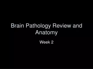
. Minimal Change GN: Synonyms: Nil disease, lipoid nephrosis, foot process disease Incidence: 80% of nephrotic syndrome in children (1-8 yrs.), mostly male. Adults in 2nd-3rd decade. Etiology: Idiopathic. Loss of net negative charge destruction of podocyte foot processes. Clinical Features: Nephrotic syndrome. History of recent URI in 30%. Association with Hodgkin’s lymphoma. Overlap with FSGS patients. Lab Features: Nephrotic urine (polyuria, Selective proteinuria. (albuminuria). Pathology: Normal Microscopy. IF - Negative. EM loss of foot processes. Clinical Course: Spontaneous remission in 25-40%. Complete remission in 65-70% of patients. Steroid resistant patients may progress to FSGS. Normal Microscopy Loss of foot process - EM
. Focal Segmental GN: Adults Synonyms: Focal segmental Sclerosis Incidence: 10 - 35% of nephrotic syndrome in adults. Etiology: Idiopathic - ? Auto Immune. No deposits. (Similar to minimal change). Clinical Features: Nephrotic syndrome. History of recent URI in 30%. Association with Hodgkin’s lymphoma. Overlap with MCD patients. Lab Features: Nephrotic urine (more, clear) Selective proteinuria. No specific laboratory findings. Podocyte damage, Segmental collapse of glom. increase in matrix (pink). Pathology: Clinical Course: Spontaneous remission 30% , 50% progression to chronic renal failure, 20% rapid progression.
. Membranous GN: Synonyms: membranous GN Incidence: 40-60 Years, 50% of adult nephrotic syndrome. Wireloop Etiology: Immune complex deposition. Idiopathic in most patients, associated with infections, drugs, carcinomas, and heavy metals. Clinical: Nephrotic syndrome in 80%, asymptomatic proteinuria in 20%. Microscopic hematuria. Lab: Non-selective proteinuria ± hematuria. Path: Diffuse, uniform BM thickening with subepithelial projections (“spikes”). Diffuse, coarsely granular IgG and C3 deposits along basement membranes. Electron-dense subepithelial deposits. Clinical Course: Excellent prognosis in children. Some adults develop ESRD. Exclusion of other diseases is required.
. Acute Post Strept, Diff, Prol GN: Synonyms: Acute proliferative glomerulonephritis, acute post-infectious GN. Incidence: Etiology: children (3-14). Sporatic, mostly winter and spring. Glomerular trapping of circulating immune complexes. (Group A, Beta-hemolytic streptococci, type 12). Clinical: Acute nephritic following strep. pharyngitis or pyoderma. (Other infections rare) Lab: Nephritic urine (little, dark, smoky) RBC casts, non selective proteinuria. Decreased serum complement. Evidence of strep inf. Path: Enlarged, hypercellular glomeruli with endothelial and mesangial cell proliferation, neutrophils, IgG and C3 in very coarsely granular pattern along GBMs. Discrete, subepithelial “hump-like” deposits. Clinical Course: Children - Excellent prognosis. Adults Worse prognosis, some develop progressive disease.
. IgA Nephropathy (Berger’s) • Commonest form of GN – Nephritic. • Young 15-30y, males, Asia-Pacific. • IgA deposits in mesangium, High serum IgA, varied severity • Episodic asymptomatic hematuria • microscopic hematuria (40%) • Bouts of macro hematuria (40%) • Nephritic or Nephrotic (rare). • Renal failure (10%) • Slowly progressive CRF in 1/3 patients. IgA dep. Normal IgA dep.
. Progression of GN: Decreased Renal reserve Renal Insufficiency Renal Failure Endstage G F R
. Acute Tubular Necrosis: • Necrosis of tubular cells – fall of as casts. • Most common cause of ARF. • Ischemic (patchy PCT & DCT) – Hypovolemia – Shock • Toxic (PCT only) – Drugs… – Toxins – Mercury, CCL4, Radiocontrast.
. Acute Tubular Necrosis(ATN): toxic Necrotic PCT (no nuclei) Glom. Norm Normal DCT (Pro. cast inside) PCT early necrosis
. “Look at the sky. We are not alone. The whole universe is friendly to us and conspires only to give the best to those who dream and work.” - Wings of Fire: An Autobiography of Dr. APJ Abdul Kalam.
. T4W3: Week overview: 2013 Term 4 CPC 3 Title: Male Genitourinary System:GU + Renal Aim: Understanding pathology ,presentation and clinical diagnosis of patients with urinary obstruction & urinary tract infections. Learning 1. Demonstrate competency in history taking & clinical outcomes: examination of patients with urinary symptoms. The student will be 2. Demonstrate competency in the clinical examination of able to the abdomen and pelvis 3. Describe the Laboratory investigations for patients with bladder outflow obstruction and haematuria 4. Demonstrate competency in debating the use of the PSA in individual patients and as a screening test. 5. Describe the first line management of prostate cancer and BPH 6. Describe the anatomy and histology of bladder, urethra + prostate;
. Week Learning outcomes: 2013 Term 4 CPC 3 Title: Male Genitourinary System:GU + Renal Aim: Understanding pathology ,presentation and clinical diagnosis of patients with urinary obstruction & urinary tract infections. Learning 7. Describe the pathology of BPH, prostate cancer, renal outcomes: and bladder tumours ,and renal and bladder calculi The student will be 8. Outline the Professional, ethical & legal issues in able to diagnosis & management of patients with benign prostatic hyperplasia (BPH) and prostate cancer 9. Outline the Epidemiology & Public Health issues of BPH and prostate cancer
. SAQ: UT Obstruction • What are differential diagnosis? • What complication he has? Or he may develop? • Should PSA be tested for all? Diagnostic levels? • When is biopsy indicated? • Does BPH lead to Carcinoma? • What is the best screening test for Ca? • What investigations are available? • BPH & Carcinoma – microscopy? • Gleason grading of prostate carcinoma?
. Self Assessment: • • • • • • Common obstructions of LUT – age Etiology, pathogenesis, morphology: BPH PSA levels in diagnosis – debate. Cystic diseases of Kidney – ADPKD Urinary Tract Infections – Microbiology. Nephrolithiasis – common stones morphology. • Transitional cell carcinoma – brief • U:C ration – significance, diagnosis.
. Core Learning Issues: (CLI) Major: • Disorders of Prostate – Prostatitis, BPH & Ca. • Nephrolithiasis: Features, Types, Pathogenesis. • Tumors of Kidney. – RCC, TCC, Wilms. • Urinary Tract Infection – Common Microbiology. Minor: • hematuria, strictures, obstructions, polyps. • Tumors of Urinary tract and bladder. • Kidney Cysts, Hydronephrosis, Recurrent UTIs, Pyelonephritis, renal abscess, Congenital disorders of kidney.
. When you lose, don’t lose the lesson! Lao Tzu Everyone makes Mistakes, only intelligent learns from it.
. Causes of Obstructive Uropathy INTRINSIC: Calculi - Lithiasis Strictures – congenital, inflammatory (UTI) Tumors – Transitional cell papilloma & Carcinoma. Blood clots, necrotic tissue (Papillary necrosis) EXTRINSIC: Pregnancy Inflammation- STI / PID, peritonitis, diverticulitis, salphingitis. Tumors: Prostate, rectum, bladder, ovaries etc.
. Nephrolithiasis: • Usually unilateral, small 1-3 mm, • Flank pain & tenderness – renal capsule. • Passage marked by Paroxysmal, intense colicky pain in the back (loin) with radiation to anterior (renal or ureteral "colic“) • “writhing in pain, pacing about, and unable to lie still” • Hematuria macro/micro • Larger stones that cannot pass produce hydronephrosis or hydroureter.
. Levels - Clinical symptoms • Ureteropelvic junction - deep flank pain No radiation. Distension of the renal capsule. (Symp. T11-L2) • Ureter – Acute, severe, colicky pain in the flank and ipsilateral lower abdomen with radiation to the testes/vulva (ilioinguinal n.). nausea / vomiting. – Upper ureter – cholecystitis. – Middle – appendicitis – Distal ureter – Pelvic Infl. Dis. • Ureterovesical junction - Cause irritative voiding, urinary frequency and dysuria. Calcium Oxalate
. Nephrolithiasis: Organic matrix(3%) + salts (97%) ~ • Calcium stones (80%): oxalate/phosphate/urate salts. Calcium Oxalate – Increased gut absorption or defective tubular reabsorphtion of calcium – Common, high pH. – Hyperparathyroidism (10%) – Hyperuricosuria – high pH • Struvite Stones (15%) magnesium ammonium phosphate (triple phos). Staghorn stone. – Chronic UTI with gram-negative rods (split urea) pH >7 – Proteus, Pseudomonas, and Klebsiella (not E. coli). • Uric acid stones (6%): – pH <5.5, high protein (meats), malignancy, 25% have gout.
. BPH-Bladder Gross – Identify Cues? Trabeculations Hypertrophy of wall Stone - urolithiasis Inflammation Median lobe- ball valve. Enlarged prostate.