Download
1 / 46
470 likes | 733 Views
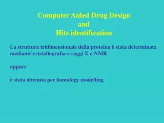
Computer Aided Drug Design and Hits identification La struttura tridimensionale della proteina è stata determinata mediante cristallografia a raggi X o NMR oppure è stata ottenuta per homology modelling. De novo drug design Building

E N D
Computer Aided Drug Design and Hits identification La struttura tridimensionale della proteina è stata determinata mediante cristallografia a raggi X o NMR oppure è stata ottenuta per homology modelling
De novo drug design Building Ricerca con gruppi funzionali partendo da un sito ed espandendosi nel sito attivo. Linking Ricerca con gruppi funzionali partendo da diversi punti del sito attivo e collegandoli successivamente con uno scaffold. In silico screening Docking da librerie virtuali o reali Scoring function
Drug Design Modification of ligands in situ or design of new ligands Docking of designed ligands into the binding site (AutoDock, Dock, FlexX…) Score of the new complexes Synthesis of new compounds
The Master Equation DG = DH - TDS
DH is relatively easy to calculate • Enthalpy (DH) is derived from static models of molecular or bimolecular structure. • Molecular mechanics force field methods deconvolve DH into intramolecular and intermolecular terms from bond stretches, angle bends, torsions, etc., electrostatic interactions and van der Waals (London) forces. • Many academic and commercial force field programs are available, using similar approaches with essentially comparable results.
DS is much harder to calculate • Entropy = disorder. Computers don’t like disorder! • Must account for all components of the system, including solvent molecules. (Explicitly?) • Must add “movement” to the molecule(s) using something like molecular dynamics. • Entropy itself is not directly (experimentally) measured – calibration of model calculations is less reliable than for enthalpy.
How can we calculate DG directly? • Free Energy Perturbation method (Kollman, Karplus, Beveridge and others) Thermodynamic cycle: DDGbind = DGI2 - DGI1 = DGenz – DGsol (DGenz = free energy binding difference between two ligands I1 and I2, DGsol = solvation energy difference between I1 and I2) DGenz and DGsol calculated from extensive molecular dynamics simulations of enzyme/inhibitor systems. • MC/MD LR (Monte Carlo/Molecular Dynamics with a Linear Response method (Jorgensen et al.) DG = b[DHelec] + a[DHvdw] + g[DSASA] 106 to 107 analyzed configurations
Metodi di predizione • Decomposizione del DG° (la teoria dice che è una procedura scorretta) • Metodi FEP (Free Energy Perturbation) [Kollman, Karplus, Beveridge….] • MC/MD LR (Monte Carlo/Molecular Dynamics con un metodo Linear Response) [Jorgensen et al.] • Metodi empirici
A “natural” force field • No preservatives and 9944/100% Hamiltonian and wave function free! • Biological binding events are not a neat set of terms specific to hydrogen bonding, acid-base, Coulombic and/or hydrophobic interactions – binding is a concerted process! • Design a Free Energy force field derived from an experiment that measures the free energy of molecular interactions.
Log PA = Log[Aoct/Awater] Log Po/w = DG° / 2.303 RT octanole water The partition of a compound between water and octanole is a process driven by intermolecular interactions between the solvent and the compound, and by hydrophobic interactions, involving solvation-desolvation. These events are the same that take place in the formation of a ligand-protein complex.
Hydrophobicity • Measured as Water / Octanol Partition Coefficient (P) • log P > 0 : lipid phase log P < 0 : water phase
Leo (CLOG-P) Method i = number of occurrences of the fragment constant f of type n. j = number of occurrences of the factor F of type m.
HINT (Hydropathic INTeraction) Software model based on experimental LogPO/W values for interaction classification andquantitative scoringevaluatesenthalpy and entropy HINT calculates empirical atom-based hydropathic parameters that encode all significant intermolecular and intramolecular non-covalent interactions implicated in drug binding or protein interactions and folding. The hydrophobic atom constants are calculated using an adaptation of the fragment constant methods of Leo and Rekker.
The “HINT equation” HINT SCORE = SS bij = SS aiSi ajSj Rij Tij + rij a = hydrophobic atom constant S = solvent accessible surface area Rij = exponential (e-r) Tij = discriminant function for polar-polar interactions rij = van der Waals term
S ai = Log Po/w = -DG / 2.303 RT SSbij = f (DG) (G. E. Kellogg and D. J. Abraham Eur. J. Med. Chem.2000, 35, 651-661)
Classes of Non-Covalent Interactions Note: Any comprehensive method that attempts to model ligand binding must also consider the energy of solvation and entropic contributions to the binding process.
Hydropathic Interactions Hydrophobic Polar Lewis Acid (H-Bond Donor) Polar Lewis Base (H-Bond Acceptor) Hydrophobic Hydrophobic Interaction Hydrophobic-Polar (desolvation) Hydrophobic-Polar (desolvation) Polar Lewis Acid (H-Bond donor) Hydrophobic-Polar (desolvation) Coulombic Repulsion Acid-Base (Hydrogen Bond) Polar Lewis Base (H-Bond Acceptor) Hydrophobic-Polar (desolvation) Acid-Base (Hydrogen Bond) Coulombic Repulsion
Correlazione tra l’energia libera di legame tra proteina e ligandi determinata mediante HINT e l’energia libera determinata mediante metodi sperimentali
Working Procedure Model Building • Starting point: protein-ligand complexes for which 3D structure (PDB files) and experimental binding affinity are determined • Hydrogen atoms added and minimized, hydrogen bound to polar atoms examined and optimized • Evaluation of the protonation state of ionizable groups on protein and ligand (Computational Titration - Fornabaio et al. J. Med. Chem. 2003, 46, 4487-4500) • Optimization of water molecules bridging protein and ligand • Hydropathic Analysis for the evaluation of all contributions to the ligand-protein complex formation
General Relationship between Hint Score and DG° 93 complexesformed by 18 different proteins of know 3D structure, R < 3.2 Å DG° = -0.0018 HINT score – 3.9041 R2 = 0.47 R = 0.68 se = 2.33 Kcal/mol Cozzini et al., J. Med. Chem. 2002, 45, 2469-2483
General Relationship between Hint Score and DG° 73 complexesR < 2.5 Å and at least 3 ligands for each protein DG° = -0.0024 HINT score – 2.2187 R2 = 0.65 R = 0.81se = 1.89 Kcal/mol Kellogg et al. J. Mol. Graph. Model. 2004, in press
Which is the contribution of water molecules bridging ligand and protein to the free energy of binding?
Active site of HIV-1 Protease in the absence and presence of a ligand
Water molecules in Ligand Binding to HIV-1 Protease D25 D125 R108 R8 R187 R87 w313bis’ w313bis w313’ w313 D29 D129 w301 I150 I50 wat301 • is crystallographically detected in the active site • occupies the same position in all complexes • usually forms four hydrogen bonds – two with the protein and two with the ligand • plays a crucial role in molecular recognition wat313, 313’, 313bis, 313bis’ • not always crystallographically detected • located in a more peripheral area of the active site
D25 D125 R108 R8 R87 R187 D29 D129 I150 I50 The software GRID (P. Goodford, J. Med. Chem. 1985) was used to locate water molecules in HIV-1 protease active site, when they were not present in the crystallographic structures.
The Role of Structural Waters in HIV-1 Protease-Ligand Complexes withoutandwithwat301 R2 = 0.30 R = 0.55 SE = 1.3 Kcal mol-1 R2 = 0.63 R = 0.80 SE = 1.0 Kcal mol-1
The Role of Structural Waters in Ligand Binding to Proteins with wat301-313 with wat301-313-313’-313bis SE = 1.0 Kcal/mol SE = 0.8 kcal/mol R = 0.77 R = 0.84 with wat301-313-313’-313bis-313bis’ with wat301-313-313’ SE = 1.0 Kcal/mol SE = 1.0 Kcal/mol R = 0.78 R = 0.78
The role of pH on Ligand Binding Penicillopepsin-Phosphonate Ligand Complex pH 3.5 pH 4.5 pH 5.5 • Binding constants experimentally determined at three different pH values(Bartlett et al., J. Org. Chem. 1990). • Models corresponding to the different protonations of the two catalytic aspartates were built, assuming that addition of one H decrease in one pH unit.
The role of pH on Ligand Binding Computational Titration • Drop protons into the molecular models, one hydrogen at a time acidification in silico of the environment of the protein-ligand complexes at the binding site • Model ALL possible cases for each “pH” level (corresponding to a defined number of protons into the model) • Calculate the HINT score for each model, averaging the values that correspond to each “pH” level • Plot the mean HINT score values as a function of the number of added protons
The Role of pH on Ligand Binding: HIV-1 Protease-Peptidic Ligand Complex For a complex between HIV-1 protease and a peptidic ligand (Glu-Asp-Leu), binding affinities were experimentally determined as a function of pH. experimental titration curve (J. M. Louis et al., Biochemistry 1998, 37, 2105-2110)
_ _ O O O O O O O O + H H H _ _ ? N N O O _ _ _ _ O O H H N N N N b b ASP25 ASP25 2 2 a a ASP29 ASP29 O O H H O O O O O O _ _ O O _ _ O O _ _ O O O O O O a a ASP25 ASP25 a a ASP30 ASP30 O O Glu-Asp-Leu bound at HIV-1 protease … … 8 ionizable groups ………. 4374 models to be evaluated
pH of crystallization How many protons should be dropped into the models? How many models should be built? How much time does it take? Which is the most favourable ionization state? All possible ionization states are modeled and scored automatically with the “COMPUTATIONAL TITRATION” procedure
Computational Titration: neuraminidase-inhibitor complexes Active site of the complex neuraminidase-DANA (2,3-didehydro-2-deoxy-N-acetylneuraminic acid) • Conserved interactions : • Three Arg interact with ligand carboxylate • The hydroxyl groups (O8, O9) of the glycerol side chain hydrogen bonded with Glu276 • The hydroxyl O4 sits at the entrance of the pocket formed by Asp151, Glu119 (Glu227)
O O A S P 1 5 1 O O A S P 1 5 1 O H H N O H H N O O O H N H 2 H O H O O O O O O H O O H O G L U 1 1 9 G L U 1 1 9 O O G L U 2 7 6 O G L U 2 7 6 O - O O - O O 2 A R G 2 9 2 A R G 2 9 A R G 1 1 8 A R G 1 1 8 A A R R G G 3 3 7 7 1 1 Computational Titration: neuraminidase-inhibitor complexes The titration curves show a peak HINT score (maximal binding energy) that should correspond to the “optimum” pH for binding.
p120GAP GTPase-activating domain Hsp90 geldanamycin-binding domain Ribonuclease A HIV-1 protease Lipid Binding Protein TOPOGRAPHIC WATER CLASSIFICATION
0-1 1-2 2-3 3-4 4-5 Total number of analyzed water molecules: 817 mean ranking: 1.6 mean HINT score PW: 199
Next: Analisi delle classi di molecole d’acqua (in cavità, in superficie, in siti attivi,…) Analisi dell’interazione tra proteina e DNA In silico screening Analisi dell’interazione proteina-proteina