Cardiovascular System
300 likes | 461 Views
Cardiovascular System. Blood. Composition of Blood. Blood is the body’s only fluid tissue It is composed of liquid plasma and formed elements Formed elements include: Erythrocytes, or red blood cells (RBCs) Leukocytes, or white blood cells (WBCs) Platelets ( Thrombocytes )

Cardiovascular System
E N D
Presentation Transcript
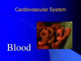
Cardiovascular System Blood
Composition of Blood • Blood is the body’s only fluid tissue • It is composed of liquid plasma and formed elements • Formed elements include: • Erythrocytes, or red blood cells (RBCs) • Leukocytes, or white blood cells (WBCs) • Platelets (Thrombocytes) • Hematocrit – the percentage of RBCs out of the total blood volume
Composition of Blood Figure 18.1
Physical Characteristics and Volume • Blood is a viscous, opaque fluid with a metallic taste. • Color varies from scarlet (oxygen-rich) to dark red (oxygen-poor) • The pH of blood is 7.35–7.45 • Temperature is 38C, slightly higher than “normal” body temperature • Average volume of blood is 5–6 L for males, and 4–5 L for females
Functions of Blood • Transports gases, nutrients, hormones, and metabolic wastes • Regulates pH and electrolyte balance • Hemostasis: maintains blood volume • Defends the body against toxins and pathogens • Stabilizes body temperature
Blood Plasma • Plasma makes up 55% of the total blood volume: • 92% of plasma is water • Proteins • Albumins: maintain osmotic pressure of blood • Globulins: antibodies and transport proteins • Fibrinogens: involved in blood clotting • Nonprotein nitrogenous substances – lactic acid, urea, creatinine • Organic nutrients – glucose, carbohydrates, amino acids • Electrolytes – sodium, potassium, calcium, chloride, bicarbonate • Respiratory gases – oxygen and carbon dioxide
Erythrocytes (RBCs) • Biconcave discs • Lack a nucleus • Filled with hemoglobin (Hb), a protein that functions in transporting Oxygen and Carbon Dioxide • Normally produced in the red marrow of bones.
Erythrocyte Function • Erythrocytes are dedicated to respiratory gas transport • Hemoglobin reversibly binds with oxygen and carbon dioxide • Hemoglobin is composed of: • The protein globin, made up of two alpha and two beta chains, each bound to a hemegroup. • Each heme group carries an atom of iron, which can bind to oxygen. • As oxygen levels decrease, hemoglobin molecules release oxygen and the globin portion binds to carbon dioxide. • Each hemoglobin molecule can transport four molecules of oxygen
Hormonal Control of Erythropoiesis • Erythropoietin (EPO) release by the kidneys is triggered by: • Hypoxia (low oxygen) due to decreased RBCs • Decreased oxygen availability (high altitude) • Increased tissue demand for oxygen (exercise) • Enhanced erythropoiesis increases the: • RBC count in circulating blood • Oxygen carrying ability of the blood increases
Hormonal Control of Erythropoiesis Figure 18.6
Leukocytes (WBCs) • Leukocytes, the only blood components that are complete cells: • Are less numerous than RBCs • Make up 1% of the total blood volume (normal count 6000-9000 per cubic mm.) • Can leave capillaries through tissue spaces via diapedesis • Leukocytosis– WBC count over 11,000 per cubic mm. • Normal response to bacterial or viral invasion
Leukocytes (WBCs) • Two categories: • Granulocytes: • Neutrophils: Phagocytic, release cytotoxic chemicals • Eosinophils: attack antibody-labeled and parasitic pathogens • Basophils: release histamines, enhance inflammation • Agranulocytes • Lymphocytes: provides specific immune response • Monocytes: macrophages engulf pathogens or debris
Platelets • Cell fragments • Functions in the clotting mechanism (Hemostasis) • Forms a temporary “Platelet Plug” that helps seal breaks in blood vessels.
White blood cells Platelets Red blood cells
Neutrophil Lymphocyte
Eosinophil Lymphocyte
Neutrophil Basophil Monocyte
6 3 4 2 5 1
6. Neutrophil 3. Eosinophil 4. Neutrophil 2. Lymphocyte 5. Basophil 1. Monocyte
Sickle cell anemia: abnormal formation of hemoglobin, which causes RBC’s to be deformed, and causes poor oxygen circulation, and blood clots.
Leukemia: Cancer of blood forming tissues, causing elevated levels of and abnormal formation of WBC’s.
Hemostasis • A series of reactions designed for stoppage of bleeding • During hemostasis, three phases occur in rapid sequence • Vascular Phase • Platelet Phase • Coagulation (blood clotting) Phase
Hemostasis • Vascular Phase: • Smooth muscles in the walls of blood vessels constrict • The decrease in the diameter of the blood vessel slows and sometimes stops blood loss. • Endothelial cells at an injury site become sticky, and cells may stick together further blocking the injury site.
Hemostasis • Platelet Phase: • Platelets attach to “sticky” endothelial cells at the injury site. • As more platelets arrive, a “Platelet Plug” forms, a mass of cells that may block the break in the vessel wall.
Hemostasis • Coagulation Phase: • Coagulation = Blood Clotting • Occurs when a series of steps results in the formation of fibrin. • Fibrin is an insoluble protein “net” that traps blood cells and platelets which form a blood clot. • The blood clot seals off the damaged portion of the blood vessel.
Blood Typing • A system of categorizing blood based on the surface antigens of RBC’s (agglutinogens) • Three of the most important antigens: A, B and Rh • When serum containing anti-A or anti-B agglutinins is added to blood, agglutination will occur between the agglutinin and the corresponding agglutinogens • Positive reactions indicate agglutination (clumping) and incompatible blood type.


