Action Potentials
Action Potentials. DR QAZI. OBJECTIVES. Define the action potential. Describe the changes during action potential. Discuss conduction (propagation) of action potential Describe recording of monophasic action potential. Action Potential.

Action Potentials
E N D
Presentation Transcript
Action Potentials DR QAZI
OBJECTIVES • Definethe action potential. • Describe the changesduring action potential. • Discuss conduction (propagation) of action potential • Describe recording of monophasic action potential.
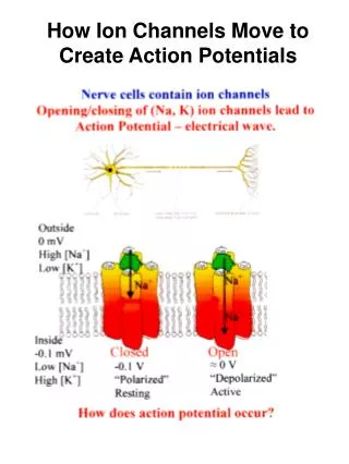
Action Potential -Rapid, large alterations in the membrane potential during which time the membrane potential may change 100 mV, ( -70 to +30), and then repolarize to its RMP Nerve Muscle cells Endocrine Immune Reproductive cells
FUNCTIONS OF AP • Relay a neuron’s message over a relatively long distance, leading to NT release. • Relay the activation signal over the surface of a muscle cell. • “Motivate” neuroendocrine cells to release hormones. • Spread an activation response over the membrane surface of immune cells. • Relay the message of fertilization over the surface of an egg.
Stimulus:sudden change of (internal or external) environmental condition - cell. 5 types 1.Submiminal 2. Miminal 3.Submaximal 4. Maximal 5. Supra maximal
Local Response • It s a graded potential • Its propagation is electronic conduction • Subthreshold stimulus • It can be summed by 2 ways • Spatial summation • Temporal summation Excitatory a d b Excitatory c Inhibitory a b Membrane Potential (mV) c d Time Temporal Summation Temporal &Spatial Summation
1. All-or-none principle. 2. Amplitude- same.
CODING OF INFORMATION 1. 2. 3. Weak stimulus Moderate stimulus Strong stimulus • Pattern = Intensity of stimulus frequency of APs • Place = type of stimulus Visual, auditory, pain, etc. • Brain area that receives signal Doctrine of Specific Nerve Energies
Stages of AP 1. NORMAL, UNPOLARIZED, EQULIBRIUM 2. POLARIZED RMP 3. STIMULUS 4. ARTIFACT 5. FIRING LEVEL 6. ELECTROTONIC POTENTIAL GP 7. THRESHOLD 8. UPSTROKE / ASCENDING WAVE /DEPOLARIZATION 9. OVERSHOOT 10. ZERO –LEVEL ISOPOTENTIAL PEAK 11. DOWN STROKE/DESCEDINGWAVE/ REPOLARIZATION 12. SPIKE POTENTIAL 13. -ve AFTER DEPOLARISATION 14 +ve AFTERDEPOLARISATION / HYPERPOLARIZATION/ UNDER SHOOT
RMP= -65mV -65
Electrotonic potentials & local response10-20 mv< -ve than the RMP . trigger AP 2 types ;- • cat –electrotonic potentials= is a depolarising---- allows the Na+ ion to move in • an–electrotonic potentials = is a hyperpolarising current allows the k+ ion to move out
+30 = 100 mV Amplitude Depolarization Na+ influx 0 - 70 mV to +30 mV -55 -70 -80 Time
Rapid depolarization Na+ + - - + Na+ Na+ • When partial depolarization reaches the activation threshold,voltage-gated sodium ion channels open. • Sodium ions rush in. • The membrane potential changes from -70mV to +40mV.
+35 Repolarization K+ efflux 0 -60 -70 -80 Time
Repolarization Na+ K+ + K+ - Na+ Na+ K+ • Na++ion channels close and become refractory. • Depolarization triggers opening of voltage-gated K+ion channels. • K+ ions rush out of the cell, repolarizing and then hyperpolarizing the membrane.
After- hyperpolarization +35 0 -60 -70 -80 Time
Refractory Period • Absolute Lasts 1 msec • Complete insensitivity exists to another stimulus • From beginning of action potential until near end of repolarization. • No matter how large the stimulus, a second action potential cannot be produced. • Has consequences for function of muscle • Relative • A stronger-than-threshold stimulus can initiate another action potential
Voltage-Gated Na++Channel 1. M gate= activation gate on Na channel; opens quickly when membrane is depolarized 2. H gate- inactivation gate on Na channel; Closes slowly after membrane is depolarized
Gate on the Delayed Rectifier K+Channel SINGLE GATE (N) that stays open as long as Vm is depolarized. slowly this allows the Vm to depolarize due to Na influx; Na and K currents do not offset each other right away
Hogkin’s cycle Positive feedback loop gradedNa+ potential Na+ enters (depolarization V-gate Na+ channels open
Action Potential Propagation: Local Currents depolarize adjacent channels causing depolarization and opening of adjacent Na channels Question: Why doesn’t the action potential travel backward?
Transmission of a signal • Think dominoes! • start the signal knock down line of dominoes by tipping 1st one trigger the signal • propagate the signal do dominoes move down the line? no, just a wave through them! • re-set the system before you can do it again, have to set up dominoes again reset the axon
Saltatory ConductionImpulse Conduction in Myelinated Neurons Most Na+ channels concentrated at nodes. No myelin present. Leakage of ions from one node to another destabilize the second leading to another action potential in the second node. And so on….
: : :