Download

1 / 71
740 likes | 1.21k Views
Revised American Thyroid Association Guidelines for the Management of Medullary Thyroid Carcinoma. Dr Zahra GhasemZadeh Endocrine Fellow Shahid Beheshti University Of Medical Science ordibehesht 1394 – April 2015. Agenda. A : Background B : Etiology of sporadic and hereditary MTC

E N D
Revised American Thyroid Association Guidelines for theManagement of Medullary Thyroid Carcinoma Dr Zahra GhasemZadeh Endocrine Fellow ShahidBeheshti University Of Medical Science ordibehesht1394 – April 2015
Agenda • A : Background • B : Etiology of sporadic and hereditary MTC • C : Clinical characteristics and relationship between genotype and phenotype • D : Direct DNA analysis to detect mutations in the RET proto oncogene • F : Secretory products of MTC • G : Morphological examination • H : The diagnosis of MTC in patients presenting with a thyroid nodule • I : Management of patients with a thyroid nodule and histological documentation of MTC • K : Management of patients with locally advanced or metastatic MTC L • L: Management of patients following an incomplete thyroidectomy and lymph node dissection
Agenda • M : Management of normal parathyroid glands resected or devascularized during surgery • N : Hormone replacement following thyroidectomy • O : Prophylactic thyroidectomy in children with hereditary MTC • Q : Management of HPTH in patients with MEN2A • R : Evaluation of patients following thyroidectomy • S : Treatment of patients with regional metastatic MTC • T :Evaluation of patients with distant metastases • U : Diagnosis and treatment of patients with clinically evident metastases • V :Systemic Therapy • W :Treatment of patients with hormonally active metastases
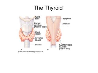
A :Background • MTC accounts for 1-2% of thyroid cancers in the United States, a much lower range than frequently cited (3-5%) primarily due to the marked increase in the relative incidence of papillary thyroid carcinoma (PTC) over the last three decades. • Over 100 years ago Jacquetdescribed a thyroid tumor with amyloid • Williams discovered that MTC originated from the neural crest • Tashjianand colleagues discovered that the C-cells secrete the Ctn • Takahashiand associates discovered the RET (RE arranged during Transfection) oncogene in 1985. • The RET protooncogene, located on chromosome 10q11.2, encodes a single-pass transmembrane receptor of the tyrosine kinase family. • RET is expressed in cells derived from the neural crest, the branchialarches, and the urogenital system
B:Etiology • all patients with MEN2A, MEN2B, and FMTC have RET germline mutations. • approximately 50% of sporadic MTCs have somatic RET mutations. • Investigators recently discovered that 18-80% of sporadic MTCs lacking somatic RET mutations have somatic mutations of HRAS, KRAS, or rarely NRAS. • The somatic RET codon M918T mutation in sporadic MTC appears to portend an aggressive clinical course and a poor prognosis. • In a recent study of 160 patients with sporadic • MTC the prevalence of somatic RET codon M918T mutations varied depending on tumor size. • <1 cm 11.3% • 1-2 cm 11.8% • 2-3 cm 31.8% • >3 cm 58.8% • the question of whether RET acts alone as the initiator of oncogenesis in sporadic MTC, or is activated later as a driver of tumor growth, other genes playing a significant role in MTC onset. • An alternate explanation for these findings is thatM918T mutated tumors have a high growth rate and are more likely to be diagnosed when they are larger
RET central to the development of sporadic and hereditary MTC • RET occur in 20-30% of patients with PTC • Activating RET translocations in patients with lung adenocarcinoma and CML. • inactivating mutations occur throughout the RET oncogene in patients with hereditary and sporadic Hirschsprung’sDisease.
At the Seventh International Workshop on MEN • North American Neuroendocrine Tumor Society, • the National Comprehensive Cancer Network, • the American Thyroid Association. • Three of the groups used either the TNM designation of the American Joint Committee on Cancer (AJCC), or terms such as Level I, II, or III, or “high”, “higher”, or “highest”, to designate progressive increases in aggressiveness of the MTC. • Recommendation 1: • The original ATA Guidelines used A, B, C, and D designations to define categories of RET mutations associated with increasing aggressiveness (from A to D) of the MTC . • D : “highest risk” (HST), RET codon M918T mutation • C : “high risk” (H), RET codon C634 mutations • A and B: “moderate risk” (MOD), RET codon mutations other than M918T and C634.Grade C
C: Clinical characteristics and relationship between genotype and phenotype 1)Sporadic MTC: • Sporadic MTC usually occurs between the 4th and 6th decades of life MTC who present with a palpable thyroid nodule • 70% of patients with have cervical metastases • 10% have distant metastases On univariate analysis prognosis is directly related to : • Patient age at diagnosis, • Male sex, • The presence of local tumor invasion, • The presence of lymph node metastases, • The presence of distant metastases. On multivariate analysis age and stage at the time of diagnosis • Ten-yearsurvival rates for patients with stages I :100% II :93% III:71% and IV :21% • The clinical behavior of sporadic MTC is unpredictable.and some patients with distant metastases may live for several years.
2)Hereditary MTC: MEN2A has expanded to include two variants: • patients with associated cutaneous lichen amyloidosis (CLA) • and patients with associated HD. • The MEN2B syndrome accounts for 5% of hereditary MTCs • Patients with MEN2B develop MTC and PHEOs and exhibit a recognizable phenotype. Strict criteria defined the diagnosis of FMTC : • more than 10 family members with MTC, • multiple carriers or affected members over 50 years of age, • and an adequate medical history (particularly in older family members) to exclude the presence of PHEO and HPTH . • A less rigid definition was the presence in at least four family members of MTC without other manifestations of MEN2A • At present, there are only 3 documented families that meet the original strict criteria described for FMTC.
Recommendation 2: There should be two MEN2 syndromes: MEN2A and MEN2B. Within MEN2A, which accounts for 95% of MEN2 cases, there should be four variants: • Classical MEN2A (represented by the uniform presence of MTC and the less frequent occurrence of PHEO, or HPTH, or both), • MEN2A with CLA, • MEN2A with HD, • and FMTC (families or individuals with RET germlinemutations who have MTC but neither PHEOs or HPTH). Grade C
2-1)Classical MEN2A • MEN2A : mutations occur in codons 609, 611, 618, or 620 of exon 10, • or codon 634 of exon 11 • all patients develop MTC and lesser numbers develop PHEOs or HPTH • the frequency of each depending on the specific RET mutation. • RET codon 634 mutations are associated with a high penetrance of PHEO and a moderate penetrance of HPTH (up to 30%). • The PHEOs are almost always benign and are usually multicentric, bilateral, and confined to the adrenal gland. • The tumors are usually associated with diffuse nodular adrenal medullaryhyperplasia, particularly in patients with RET germlinemutations in codons 918 and 634. • Patients with MEN2A and a unilateral PHEO usually develop a contralateral PHEO within 10 years.
Classical MEN2A • The HPTH in patients with Classical MEN2A is usually mild and associated with few if any symptoms. • From 1 to 4 parathyroid glands may be enlarged. • There are very rare families with features of Classical MEN2A who have no identifiable RET germlinemutation. • In this situation the diagnosis of Classical MEN2A can be made if one or more first-degree relatives have characteristic clinical features of the entity.
2-2) MEN2A and cutaneous lichen amyloidosis • Cutaneous lichen amyloidosis is a rare disorder that usually occurs sporadically but may present in a hereditary pattern. • The CLA in MEN2A is characterized by dermatological lesions that are particularly evident in the scapular region of the back corresponding to dermatomes T2-T6. • Theclassic symptom of CLA is intense pruritus that improves with sun exposure and worsens during periods of stress. • Hyperpigmentedlesions develop later, apparently secondary to scratching. • The inciting lesion appears to be notalgiaparaesthetica, a sensory neuropathy involving the dorsal spinal nerves. • The CLA may be present at a young age and prior to the onset of clinically evident MTC, thus serving as a precursor for the syndrome. • almost exclusively in patients with the RET codon 634 mutation, although it has been reported in a patient with a codon 804 mutation. • Treatment :local corticosteroid ,antihistamine ,phototherapy ,tyrosine kinase inhibitor.
2-3) MEN2A and Hirschsprung’s Disease • RET germline mutations are present in 50% of patients with hereditary HD and in 15-20% of patients with sporadic HD. • The RET mutations point mutations involving codons in exon 10: 609 (15%), 611 (5%), 618 (30%) and 620 (50%) • HD occurs in approximately 7% of patients with MEN2A • 2-5% of patients with HD have MEN2A • it is important to exclude HD in older patients with MEN2A and exon 10 RET mutations.Conversely, patients with HD who have exon 10 RET mutations should be evaluated for MEN2A.
2-4)FMTC • Familial medullary thyroid carcinoma is characterized by the presence of a RET germlinemutation in families with MTC, or single individuals with MTC but no known family history of MTC, who develop neither PHEOs or HPTH. 2-5)MEN2B • In patients with MEN2B the MTC often presents in infancy and is highly aggressive metastasizing early to regional lymph nodes and beyond • 75% of MEN2B cases are sporadic ,de novo RET mutations, • 25% cases occur in families with previous or current manifestations of MEN2B. • 95% RET germline mutations in exon 16 (codon M918T) • <5% have RET germline mutations in exon 15 (codon A883F)
MEN2B • Approximately 50% of patients with MEN2B develop PHEO • typical facies, ophthalmologic abnormalitiess • generalized ganglioneuromatosis • abdominal symptoms characterized by bloating, intermittent constipation, and diarrhea, and some patients require surgery for intestinal obstruction. • In 5-9% of patients with MEN2A, and the large majority of patients with MEN2B, the RET mutation arises de novo and almost always from the paternal allele.
D : Direct DNA analysis to detect mutations in the RET protooncogene Recommendation 6: Patients with presumed sporadic MTC should have genetic counseling and genetic testing to detect a RET germline mutation.Grade B Recommendation 7: Genetic counseling and genetic testing for RET germline mutations should be offered to: • First degree relatives of patients with proven hereditary MTC, • Parents whose infants or young children have the classic phenotype ofMEN2B, • Patients with CLA, and Infants or young children with HD and exon 10 RET germline mutations and adults with MEN2A and exon 10 mutations who have symptoms suggestive of HD. Grade B Recommendation 9 : In very rare families who meet the clinical criteria for MEN2A or 2B, despite negative sequencing of the entire RET coding region, the relatives at-risk should be periodically screened by conventional methods for MTC, PHEO, and HPTH. After the initial evaluation screening should continue at 1– 3 year intervals. Grade C
it is possible to perform prenatal testing on fetal blood cells obtained from maternal blood ,genotype analysis can detect RET mutations in the serum of patients with MEN2A. • Such testing might be offered to couples where only the father has hereditary MTC and the parents wish to know the mutational status of the fetus. • Pre-implantation genetic diagnosis (PGD) is an in vitro fertilization technique that isolates embryonic cells for single-site RET testing. The procedure involves removal of one or two cells from an 8-cell blastocyst and then sequence analysis of the DNA of the removed cells to determine if a RET mutation is present.
F: Secretory products of MTC • Calcitonin: • Calcitonin is a 32 amino acid monomeric peptide • ICMAs that are highly sensitive and specific for monomeric Ctn elevations of calcitonin : • Sepsis or other general inflammatory conditions • Chronic renal failure • HPTH, • Autoimmune thyroiditis, • Small cell and large cell lung cancers, • Prostate cancer, • Mastocytosis,and various enteric and pulmonary neuroendocrine tumors • Heterophilic antibodies • ‘‘Hook effect’’ • The serum Ctn levels in non-thyroid malignancies do not increase in response to calcium or pentagastrinstimulation,and compared to MTC the tumors usually produce less Ctnper gram of tissue.
Calcitonin: • 95th percentile for serum Ctn levels to be 5.2ng/L in women 11.7 ng/L in men • The current revised MTC guidelines do not specify reference ranges of basal or stimulated serum Ctnlevels • High procalcitonin to Ctn ratio was correlated with an increased risk of progressive disease and a shortened progression-free survival (PFS), potentially useful in predicting prognosis. • Serum Ctn concentrations are particularly high during the first week of life and in low-birth weight children and premature infants. <4.5 <75 ng /dl <6m <40 ng/dl 6m-3 y <15 ng/dl provocative testing : • In determining the timing of thyroidectomy in children who have inherited a mutated RET. • In the evaluation of patients for persistent or recurrent MTC following thyroidectomy. • Detecting MTC in patients with nodular goiters.
Carcinoembryonic Antigen • for evaluating disease progression in patients with clinically evident MTC and for monitoring patients following thyroidectomy. • False elevation CEA heterophilic antibodies, smoking tobacco, • Or other conditions, such as gastrointestinal tract inflammatory disease, benign lung disease, or a host of non-thyroid malignancies. • elevations of serum CEA and Ctn levels indicate disease progression. • increasing serum CEA levels associated with stable or declining serum Ctn levels in poorly differentiated. • The most aggressive tumors had persistent and intense CEA staining but minimal if any Ctn staining. • It was suggested that CEA is a marker for early epithelial differentiation and therefore retained, while Ctn is a late marker for terminal differentiation and therefore lost . • Rarely, patients with advanced MTC have normal or low serum levels of Ctn and CEA. which case it conveys a poor prognosis.
Recommendation13: Clinicians should be aware that falsely high or low serum Ctn levels might occur with a variety of clinical diseases other than MTC, and consider this this possibility when serum Ctnlevels are disproportionate to the expected clinical findings. Grade C Recommendation 14: In interpreting serum Ctn data, clinicians should be aware that Ctn levels are markedly elevated in children under 3 years of age, especially under 6 months of age. Also, Ctn levels are higher in males compared to females. Grade B Recommendation 15: Basal levels of serum Ctn and CEA should be measured concurrently. In patients with advanced MTC a marked elevation in the serum CEA level out of proportion to a lower serum Ctn level, or normal or low levels of both serum Ctn and CEA, indicate poorly differentiated MTC. Grade B
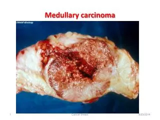
G : Morphologic Examination • On histological examination the MTC-cells are typically round, polyhedral or spindle shaped, and form sheets or nests with peripheral palisading in a vascular stroma. • The amyloid material present in over half of MTCs is actually composed of full-length Ctn. • It can be confused with PTC, FTC, paraganglioma, and lymphoma or sarcoma • The MTC cells express cytokeratins, mainly CK7 and CK18, NKX2.1 (TTF1), and chromograninA, but the most important diagnostic markers are Ctn and CEA.
PTC or FTC occurs concurrently with MTC • Their coexistence represents a coincidental collision with intermingling of neoplastic C-cells and follicular cells. • A recent study from Germany identified simultaneous MTC and PTC in 26 (2.6%) of 1019 PTCs, in 6 (2.6%) of 235 hereditary MTCs, and in 20 (4.1%) of 492 sporadic MTCs. • CCH • C- Cell hyperplasia is actually a misnomer. Considering its multicentricity, the entity most likely represents clonal proliferation of multiple transformed progenitor C-cells. Therefore, terms such as C-cell carcinoma in situ, or C-cell neoplasia are more appropriate.
The criteria for the diagnosis of CCH : • there are greater than 7 C-cells per cluster, • complete follicles surrounded by C-cells, • distribution of C-cells beyond the normal anatomical location • The CCH that occurs secondarily in association with hyperparathyroidism, chronic lymphocytic thyroiditis ,renal insufficiency, and aging, is not a premalignant condition. The distinction of CCH from microcarcinoma(MTC less than 1 cm and without capsular invasion) is challenging but clinically significant. Recommendation 16 : The assessment of a thyroid tumor with any feature suggestive of MTC should include IHC analysis to determine the presence markers such as Ctn, chromogranin, and CEA, and the absence of thyroglobulin. Grade B
H : The diagnosis of MTC in patients presenting with a thyroid nodule 1) FNA • Epithelioid Tumors : Follicular lesion • Plasmacytoid tumors : Plasmacytomas • Spindle cell tumors : Sarcomas • Amyloid : Not diagnostic Although in a meta-analysis of 15 publications the accuracy of FNA in diagnosing MTC in patients with MTC nodules was less than 50%. Recommendation 19: Thyroid nodules that are 1 cm or greater in size should be evaluated by FNA. FNA findings that are inconclusive or suggestive of MTC should have calcitonin measured in the FNA washout fluid and IHC staining of the FNA sample to detect the presence of markers such as Ctn, chromogranin, and CEA, and the absence of thyroglobulin. Grade B
2) Ctnlevel • measured serum Ctn levels in 10,864 patients with nodular thyroid disease and detected MTC in 0.40% (0.3-1.4) • serum Ctn measurements had a higher diagnostic sensitivity and specificity compared to FNA finding. • MTC is present in only 0.3–1.4% of patients with thyroid nodules , routine serum Ctnmeasurement in this population has raised concerns of cost-effectiveness, especially when many of the operated patients would have no MTC based on imperfect specificity if a cut-off was chosen that optimized sensitivity. • Ctn measurement of thyroid nodule FNA washings may significantly improve the sensitivity of the test, questions of cost effectivenessare likely to remain.
An elevated level of serum calcitonin is a highly sensitive marker for MTC, but it is not especially specific. • Only 10% to 40% of all patients with thyroid nodules associated with high basal levels of calcitonin also had MTC. • Therefore, in the remaining 60% to 90% of patients, elevated calcitonin values are secondary to other conditions
CEA, procalcitonin, CT stimulation, CT in washout of nodule ’ s aspiration
Recommendation 20: Realizing that opinions of experts vary regarding the usefulness of measuring serum Ctn levels in patients with nodular goiters, the Task Force recommends that physicians decide whether the technique is useful in the management of patients in their clinic. Grade I I : Management of patients with a thyroid nodule and histological documentation of MTC 1) Preoperative image studies If hereditary MTC or bilateral MTC is evident, PHEOs and HPTH should be excluded. The presence of bilateral MTC does not necessarily assure that the tumor is hereditary since the frequency of disease in both thyroid lobes ranges from 0-9% in patients with sporadic MTC who have no RET germlinemutations
Recommendation 21: Patients presenting with a thyroid nodule and a cytological or histological diagnosis of MTC should have a physical examination, determination of serum levels of Ctn and CEA, and genetic testing for a RET germline mutation. The presence of a PHEO and HPTH should be excluded in patients with hereditary MTC. Recommendation 22: Ultrasound examination of the neck should be performed in all patients with MTC. Contrast enhanced CT of the neck and chest, three-phase contrast-enhanced multi-detector liver CT, or contrast-enhanced MRI of the liver, and axial MRI and bone scintigraphy are recommended in patients with extensive neck disease and signs or symptoms of regional or distant metastases , and in all patients with a serum Ctn level greater than 500 pg/mL.Grade C
Patients with unilateral intrathyroidaltumors had • lymph nodemetastasesin 81% of central compartment (level VI) dissections, • 81%of ipsilateral lateral compartment (levels II to V) dissections, • 44% of contralateral lateral compartment (levels II to V) dissections • In patients with bilateral intrathyroidaltumors, nodal metastases were present in 78% of central compartment dissections, • 71% of lateral compartment dissections ipsilateral to the largest intrathyroidtumor, • and 49% of lateral compartment dissections contralateral to the largest thyroid tumor Surprisingly, the frequency of lymph node metastases in the central and ipsilateral compartments ranged from 50% to 75%, whether the primary tumors was less than 1 cm Or greater than 4 cm.
Tumors located in the upper thyroid pole metastasize first to the upper portion of the ipsilateral lateral compartment whereas tumors in the middle and lower portions of the gland spread first to the central compartment. • Apart from the location of the primary intrathyroidaltumor, the overall incidence of lateral compartment lymph node metastases is related to the frequency of central compartment lymph node metastases. • With ipsilateral lymphatic drainage of tumor cells (i.e., involvement of only the central and lateral cervical lymph node compartments on the side of the primary neoplasm), surgical cure may be attainable in some patients, whereas metastases in the contralateral lateral compartment herald incurable disease.
The preoperative basal serum Ctn level is also useful in determining the extent of lymph node Metastases Basal serum Ctn levels > 20 pg/mL, ipsilateral central > 50 pg/mL, ipsilateral lateral >200 pg/mL, contralateral central and >500 pg/mL contralateral lateral lymph node Bilateral compartment-oriented neck dissection achieved postoperative biochemical cure in at least half of the patients with pretreatment basal Ctnlevels of 1,000 pg/mL or less,but not in patients with levels greater than 10,000 pg/mL.
Recommendation 24: Patients with MTC and no evidence of neck lymph node metastases by US examination and no evidence of distant metastases should have a total thyroidectomy and dissection of the lymph nodes in the central compartment (level VI). Grade B. Recommendation 25: In patients with MTC and no evidence of neck metastases on US, and no distant metastases, dissection of lymph nodes in the lateral compartments (levels II-V) may be considered based on serum Ctn levels. The Task Force did not achieve consensus on this recommendation. Grade I (Ctn>20pg/ml) • Clearance of lymph nodes in the lateral neck is not without complications, primarily lymphatic leakage, occurring in 0.5 to 8% of patients, and damage to a spinal accessory nerve with resulting shoulder dysfunction, occurring in 25-50% of patients.
Recommendation 26: Patients with MTC confined to the neck and cervical lymph nodes should have a total thyroidectomy, dissection of the central lymph node compartment (level VI), and resection of the involved lateral neck compartments (level II-V). When preoperative imaging is positive in the ipsilateral lateral neck compartment but negative in the contralateral neck compartment, contralateral neck dissection should be considered if the basal calcitonin level is > 200 pg/mL.Grade: C • In another study metastases to 10 or more lymph nodes, or involvement of more than two lymph node compartments, precluded normalization of serum Ctn • Unfortunately, most patients with MTC and regional lymph node metastases have systemic disease and are not cured by total thyroidectomy and bilateral neck dissection.
K: Management of patients with locally advanced or metastatic MTC Recommendation 27 In the presence of extensive regional or metastatic disease less aggressive surgery in the central and lateral neck may be appropriate to preserve speech, swallowing, parathyroid function, and shoulder mobility. External beam radiotherapy (EBRT), systemic medical therapy, and other nonsurgical therapies should be considered to achieve local tumor control. Grade C L : Management of patients following an incomplete thyroidectomy and lymph node dissection Recommendation 28 Following unilateral thyroidectomy for presumed sporadic MTC completion thyroidectomy is recommended • in patients with a RET germline mutation, • an elevated postoperative serum Ctn level, • or imaging studies indicating residual MTC. • The presence of an enlarged lymph node in association with a normal serum Ctn level is not an indication for repeat surgery. Grade B
Recommendation 29In patients having an inadequate lymph node dissection at the initial thyroidectomy a repeat operation, including compartment oriented lymph node dissection, should be considered if The preoperative basal serum CTN level is less than 1,000 pg/mL and 5 or fewer metastatic lymph nodes were removed at the initial surgery. Grade C. If more than 5 metastatic lymph nodes were resected at prior surgery the biochemical cure rate fell to 5% (2 of 43 patients). When preoperative basal serum Ctnlevels exceeded 1,000 pg/mL (reference range less than10 pg/mL) biochemical cure was exceptional. M: Management of normal parathyroid glands resected or devascularized during surgery
Recommendation 30 During a total thyroidectomy for MTC normal parathyroid glands should be preserved in situ on a vascular pedicle. If all normal parathyroid glands are resected, or if none appear viable at the termination of the procedure, slivers of a parathyroid gland should be transplanted into the SCM in patients with sporadic MTC, MEN2B, or MEN2A and a RET mutation rarely associated with HPTH. In patients with MEN2A and a RET mutation associated with a high incidence of HPTH the parathyroid tissue should be transplanted in a heterotopic muscle bed. N: Hormone replacement following thyroidectomy Recommendation 31 Serum TSH should be measured within 4–6 weeks postoperatively. Replacement therapy with levothyroxine should be administered with the goal of maintaining serum TSH levels in the euthyroid range. Grade B
O: Prophylactic thyroidectomy in children with hereditary MTC Two groups of patients with MEN2B should be considered separately: < 25% who have known hereditary MEN2B and >75% who have de novo RET mutations and phenotypically normal parents. Recommendation 34: Children in the ATA-HST category with a RET codon M918T mutation should have a thyroidectomy in the first year of life, perhaps even in the first months of life. In the absence of suspicious lymph nodes the performance of a central neck dissection should be based on whether the parathyroid glands can be identified and left in situ or autotransplanted. The surgeon and pediatrician caring for the patient, in consultation with the child’s parents should decide the timing of thyroidectomy. Grade C
In children with de novo RET codon M918T mutations the diagnosis of MEN2B is usually made upon detection of a thyroid nodule. • It is critically important that physicians be aware of the characteristic phenotype associated with MEN2B, since it is almost always evident prior to the detection of a thyroid nodule or a PHEO.(skeletal ,oral ,ocular ,ganglioneuromatosis) • Children with MEN2A and RET codon 634 mutations (ATA-H category) often develop MTC during the first years of life, therefore, annual physical examination, cervical US, and measurement of serum Ctn levels should begin at 3 years of age.
Recommendation 35 • Children in the ATA-H category should have a thyroidectomy performed at age 5 years, or earlier based on the detection of elevated serum Ctn levels. A central neck dissection should be performed in children with serum Ctn levels above 40 pg/mL, or with evidence on imaging or direct observation of lymph node metastases. The surgeon and pediatrician caring for the patient, in consultation with the child’s parents should decide the timing of thyroidectomy. Grade B
Recommendation36 • Children in the ATA-MOD category should have a physical examination, US of the neck, and measurement of serum Ctn levels beginning around 5 years of age. • The timing of thyroidectomy should be based on the detection of an elevated serum Ctn level; however, 6-month or annual evaluations may extend to several years or decades. • Parents who are concerned about a long-term evaluation program, may opt to have their child’s thyroid gland removed around 5 years of age. The surgeon and pediatrician caring for the patient, in consultation with the child’s parents should decide the timing of thyroidectomy. Grade B
P: Management of PHEO in Patients with MEN2A and MEN2B: Recommendation37 • Screening for PHEO should begin by age 11 years for children in the ATA-H and ATA-HST categories and by age 16 years in children in the ATA-MOD category. • Screening consists of measuring free plasma metanephrines and normetanephrines, or 24-hour urinary metanephrinesand normetanephrines. • Adrenal imaging with CT or MRI is indicated in patients with positive biochemical results. Grade C
Recommendation 38 Patients with MEN2A or MEN2B and a histological diagnosis of MTC regardless of age and presenting symptoms must have a PHEO excluded prior to any interventional procedure. Thepresence of a PHEO must be excluded in women with MEN2A or MEN2B who are planning a pregnancy or are pregnant. If a PHEO is detected it should be treated preferably during pregnancy. Grade C Recommendation39: • If they coexist, a PHEO should be removed prior to surgery for either MTC or HPTH. Grade B Recommendation 40 After appropriate preoperative preparation a PHEO should be resected by laparoscopic or retroperitoneoscopicadrenalectomy. Subtotal adrenalectomy to preserve adrenal cortical function should be considered as an alternative procedure. Grade B