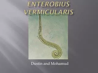
Enterobius vermicularis
Enterobius vermicularis. Presented by Sandra Thorbus & Samantha Todd. Hosts. Definitive Host: Humans Not associated with socioeconomic status. Geographical Distribution. Worldwide Most common worm infection in the United States & Western Europe.

Enterobius vermicularis
E N D
Presentation Transcript
Enterobiusvermicularis Presented by Sandra Thorbus & Samantha Todd
Hosts • Definitive Host: Humans • Not associated with socioeconomic status
Geographical Distribution • Worldwide • Most common worm infection in the United States & Western Europe. • In the United States, a study by the CDC reported incidence rate of 11.4% among all ages, 33% among children. • Prevalence in children as high as 61% in India, 50% in England, 39% in Thailand, 37% in Sweden, and 29% in Denmark.
Morphology • Egg has five membranes • Eggs are translucent and are covered in a material that allows them to stick to environmental objects • Membrane makes the eggs “itchy” • Eggs have a thick shell that is flattened on one side • The eggs small size (50-60 micrometers) • May contain an embryo or fully-developed larva • Larvae molt twice before hatching
Larvae Morphology Larvae grow to 140-150 micrometers in length Larvae are smaller and more coiled than the adults Molt twice before becoming adults
Adult Morphology White, small roundworm with cylindrical body surrounded by 3 layered cuticle The female has a sharply pointed posterior end (8-13 mm long x 0.5 mm thick) Have alae on anterior part of body wall Can lay up to 15.000 eggs/day Male has a curled posterior end measuring 2 to 5 mm long x 0.2 mm thick Both sexes have three lips
Transmission Human-to-human contact by ingesting infectious eggs Eggs remain viable in moist environment for up to three weeks Once eggs are deposited near anus they can contaminate other surfaces such as: Fingernails Hands Clothing and bed linens Then onto food, water, furniture, toys, bathroom fixtures and pets.
Prevention • Wash hands after using the bathroom and before preparing food. • Wash bedding and underclothing frequently, especially those of any affected family members.
Life Cycle Eggs are ingested Hatch in S.I. Larvae emerge and migrate through small intestine to colon Molt twice to become adults Gravid female attaches to intestinal mucosa in ileum, cecum, appendix or ascending colon Ingest colon contents until entire body is filled with eggs
Life Cycle Gravid females migrate through the colon towards the rectum at a rate of 12-14 cm/hour Female emerges from anus and deposits eggs by contracting and expelling, dying and degrading, or bodily rupture due to the host scratching. The female comes out of the anus so the eggs can be exposed to oxygen to mature After laying eggs, the female becomes opaque and dies. Under optimal conditions larvae within the eggs will develop within 4-6 hrs. after being laid making them extremely infective
Pathogenesis • Causes Enterobiasis • Retroinfection-larvae migrate back up the bowel to the G.I. tract • Pinworms as seen in colonoscopy
Symptoms • Intense itching in the anal region, especially at night • Restless sleep • Infection can migrate to the vagina and cause vaginal discharge • Itching leads to secondary bacterial skin infection • Abdominal pain and nausea are associated with high population • Some are asymptomatic
Diagnosis • Itching around perianal region is indicative of infection • Worms are visible in the anal region, especially 2 to 3 hours after sleep, • Look like tiny pieces of white thread • Eggs are rarely seen in stool samples
“Scotch Tape Test” Most reliable method to detect eggs Piece of cellophane tape is placed sticky side down to the skin around the anus Pinworm eggs will stick to the tape and then the tape can be viewed under a microscope Test should be done immediately after the person wakes up in the morning before washing, going to the bathroom, or getting dressed since eggs may be removed during these processes.
Treatment Mebendazole or Albendazole commonly prescribed for entire family OTC PyrantelPamoate 1 dose is given immediately, then wait two weeks for another dose. The second dose is to kill any adult worms that may have hatched in the meantime
Review • How many times do pinworms molt? • What is the tell tale sign of a pinworm infection? • What is one method of diagnosis? • Where do females lay their eggs? • How can you prevent pinworm infection?
Works Cited • http://www.ncbi.nlm.nih.gov/pubmedhealth/PMH0002137/ • http://en.wikipedia.org/wiki/Enterobius • http://animaldiversity.ummz.umich.edu/site/accounts/information/Enterobius_vermicularis.html