Download
1 / 97
1.21k likes | 2.19k Views
Chronic Kidney Disease (CKD). Key Concepts Chronic kidney disease (CKD) is classified based on: * the cause of kidney disease, assessment of glomerular filtration rate, and extent of proteinuria .

E N D
Chronic Kidney Disease (CKD)
Key Concepts • Chronic kidney disease (CKD) is classified based on: * the cause of kidney disease, assessment of glomerular filtration rate, and extent of proteinuria. • Frequent complications of advanced CKD include altered sodium and water balance, hyperkalemia, metabolic acidosis, anemia, CKD-related mineral and bone disorder (CKD-MBD), and cardiovascular disease. 3. Key mechanisms responsible for the progression of CKD: Reduction of kidney mass, development of glomerular hypertension, and intratubularproteinuria 4. Anemia of CKD is primarily caused by a deficiency in the production of endogenous erythropoietin by the kidney with iron deficiency as a contributing factor. 5. CKD-MBD includes abnormalities in parathyroid hormone (PTH), calcium, phosphorus, the calcium–phosphorus product, vitamin D, bone turnover, and soft-tissue calcifications and contributes to extravascular calcifications.
Guidelines provide information to assist healthcare providers in clinical decisions and the design of appropriate therapy to manage CKD progression and the associated complications. • Patient education plays a critical role in the appropriate management of patients with CKD and related complications. • ACEIs and ARBs are key pharmacologic treatments of CKD because of their effects on renal hemodynamics and reduction ofBP, which help to limit kidney disease progression. • Management of anemia includes administration of erythropoietic-stimulating agents (ESAs) (epoetinalfa, darbepoetinalfa) and regular iron supplementation (oral or IV administration) to maintain hemoglobin and prevent the need for blood transfusions. There is evidence indicating a higher risk of cardiovascular events when hemoglobin is targeted to greater than 11 g/dL. 10. Management of CKD-MBD includes dietary phosphorus restriction, phosphate-binding agents, vitamin D supplementation, and calcimimetic therapy.
CKD Definition: **Abnormalities in kidney structure or function for ≥ 3 months, with implications for health. • Structural abnormalities include: *Albuminuria of more than 30 mg/day, *Presence of hematuria or red cell casts in urine sediment, *Electrolyte & other abnormalities due to tubular disorders, *Abnormalities detected by histology, *Structural abnormalities detected by imaging, Or *history of kidney transplantation. ** An abnormality in kidney function is usually indicated by a ↓ (GFR).
The prognosis of CKD can vary and depends on: • Cause of kidney disease; (b) GFR at time of diagnosis; (c) Degree of albuminuria; (d) Presence of other comorbid conditions. *Frequent Complications of Advanced CKD: • Altered sodium and water balance, • Hyperkalemia, • Metabolic acidosis, • Anemia, • CKD-related mineral and bone disorder (CKD-MBD), • Cardiovascular disease (CVD).
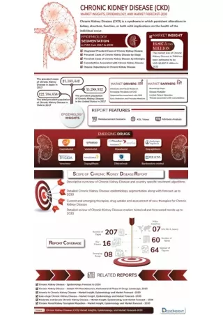
Epidemiology: • 13% of the U.S. population. • CKD is more likely in: * Those over 60 years of age * Diabetics * Hypertension patients * CVD patients • Rates of ESRD are > in African Americans (3.4 X greater) and Native Americans (0.5 X greater) compared with whites and 1.5 times > in Hispanics than in non-Hispanics.
Etiology of CKD: 1. Susceptibility Factors: • Most of these susceptibility factors are not amenable to pharmacologic or lifestyle, interventions, but are useful for identifying individuals at high risk of CKD. 2. Initiation Factors: • Conditions that directly result in kidney damage & are modifiable by pharmacologic therapy. *Diabetes mellitus: leading cause of CKD & of ESRD in the USA *Hypertension: 2nd leading cause of ESRD *Glomerulonephritis: 3rd leading cause of ESRD
3. Progression Factors: • Result in faster decline in kidney function & cause worsening of CKD. * May be modified by pharmacologic therapy or lifestyle modifications to slow the progression of CKD.
Progression Factors: • Proteinuria: • Marker of glomerular & tubular dysfunction • Degree of proteinuria correlates with progression of CKD. • Microalbuminuria> 30mg/day: linked with vascular injury & ↑ CV mortality
2) Elevated BP * Systemic BP correlates with glomerular pressure • Elevations in both systemic BP & glomerular pressure contribute to glomerular damage • The rate of GFR decline is related to elevated systolic BP
3) Elevated Blood Glucose: • The reaction between glucose & protein in blood produces advanced glycation end products (AGEs), which are metabolized in the proximal tubules. • Hyperglycemia→↑ AGEs synthesis in diabetics (suspected to cause Diabetic kidney disease)
4) Tobacco Smoking: • Induces thickening & hyperplasia of glomerulus • Raises systemic BP • Independent risk factor in developing microalbumiuria in HTN • Independnt & dose-dependent risk factor in developing CKD & microalbuminuria, & progression to ESKD. • Risk is more pronounced in men than in women
Pathophysiology of CKD: 1) Initial kidney damage from initiation factor 2)↓ # functioning nephrons 3) Remaining nephrons hypertrophy to compensate which initially may be adaptive 4) Over time →development of intraglomerular HTN may be mediated by angiotensin II (potent vasoconstrictor of both afferent and efferent arterioles, preferentially affects the efferent arterioles). Angiotensin II may also mediate CKD progression through nonhemodynamiceffects 5) High intraglomerular capillary pressure impairs the size- selective function of the glomerular permeability barrier → ↑urinary excretion of albumin and proteinuria.
Pathophysiology of CKD (cont.): 6) Proteinuria alone may → progressive loss of nephrons by direct cellular damage. (Filtered proteins i.e., albumin, transferrin, complement factors, IGs, cytokines, & angiotensin II in the renal tubule → ↑production of inflammatory & vasoactive cytokines (i.e. endothelinand monocyte chemoattractant protein-1 (MCP-1) → tubular cell toxicity. 7) Proteinuria may also →activation of complement components on the apical membrane of proximal tubules which may be the key mechanism of damage in the progressive proteinuricnephropathies. 8) These events ultimately → a) scarring of the interstitium, b) progressive loss of structural nephron units, c) reduction in GFR.
Assessment: • Screening for CKD should be done in all pts. with↑’d risk for developing CKD. • Assessment for CKD should include: *SCr measurement *Urinalysis *BP *Serum electrolytes &/or*Imaging studies
Proteinuria is the 1⁰ marker for structural kidney damage even with normal GFR. • Clinically significant proteinuria is defined as: * urinary protein execret’n > 300mg/d Or *Spot urine dipstick > 30 mg/dL Microalbuminuria: 30-300 mg urinary albumin/day
Clinical Presentation: • Symptoms • Stages 1& 2 CKD are generally asymptomatic • Stages 3& 4 may be associated with min. symptoms • Stage 5 can be associated with Uremic symptoms (fatigue, weakness, shortness of breath, mental confusion, nausea and vomiting, bleeding, and loss of appetite), itching, cold intolerance, weight gain (from accumulation of fluid), and peripheral neuropathies • Signs: • CV: Worsening HTN, edema, dyslipidemia, LVH, ECG changes, CHF • Muscular: Cramping • Neuropsychiatric: Depression, anxiety, impaired mental cognition • GI: GERD, GI bleeding, abdominal distension • GU: Changes in urine volume & consistency, "foaming" of urine (indicative of proteinuria)
Laboratory tests • All Stages 1-5 CKD: ↑BUN, SCr, ↓GFR • Advanced Stages: • Decreased: bicarbonate (metabolic acidosis), RBCs/Hb/Hct (anemia), iron indices (iron deficiency), vitamin D levels, albumin (malnutrition), glucose (may result from decreased degradation of insulin with impaired kidney function or poor oral intake), and calcium (in early stages). • Increased: potassium, phosphorus, magnesium, PTH, HTN, glucose (uncontrolled diabetes is a cause of CKD), LDL and TG, and calcium (in ESRD). • Other: may be hemoccult-positive if GI bleeding occurs (uremia) Urine may be positive for protein • Other diagnostic tests: Structural abnormalities of kidney may be present on diagnotic test
Treatment: • The 1⁰ goal : To slow & prevent the progression of CKD. • Therefore, it requires early identificat’n of pts @ risk for CKD to initiate interventions early in the course of the dz.
Nonpharmacologic Therapy: • Nutritional Management: • ↓dietary protein intake → slows the progression of kidney dz. • Recommendation: • GFR< 25 ml/min→↓ protein to 0.6g/kg/d • No adequate dietary energy intake → ↑ protein to 0.75 g/kg/d
Malnutrition: common in CKD & may be due to: *↓’d appetite * hypercatabolism * nutrients losses through dialysis • Dialysis CKD pts → 1.2-1.3 g/kg/d • Recommendation: • DM pts with CKD stages 1-4 → limit protein to 0.8 g/kg/d **↓ salt intake to < 2 g/day of sodium ( 5 gNaCl) with hypertension or proteinuria.
Pharmacologic Therapy: • Intensive Blood Glucose Control in DM Pts: • Target HbA1c < 7% to ↓proteinuria with or without diabetic KD • Intensive insulin therapy was effective in delaying development & progression of diabetic KD in types 1 & 2 DM: Insulin 3 or more X/day to maintain preprandial BG 70-120 g/dL & postprandial BG < 180 g/dL
2) Optimal BP Control: Recommendation: ** Goal BP < 130/80 mmHg in CKD <125/75 mmHg in proteinuria • All antihypertensives have similar effects on BP ** ≥ 3 agents are required to achieve the BP < 130/80
3) Reduction of Proteinuria: *ACEIs & ARBs ↓ glomerular capillary pressure & volume due to their effects on Angiotensin II → ↓proteinuria (independent of their ↓ BP) ** ACEIs & ARBs: Antihypertensives of choice for all CKD pts unless contraindicated due to their ability to ↓ proteinuria more than any other antihypertensives (up to 35-40%) • Nonhydropyridine CCBs also↓proteinuria (related to their effects on BP↓)
4) Hyperlipidemia: ** Goals of Tx of dyslipidemia are to: 1st to ↓ atherosclerotic CVD risk 2nd to ↓ proteinuria & decline in kidney function in CKD pts.( prteinuria is associated with ↑d TC, LDL-C, TG) **Statins & Fibric acid derivatives (NOT combined due to ↑d rhabdomyolysis ): First-line therapy unless contraindicated. ** Hyperlipidemia Tx can ↓ proteinuria & CKD progression. **Most statins are affected by enzyme inhibitors EXCEPT Pravastatin & Fluvastatin
5) Smoking Cessation & Exercise: *Smoking cessation is encouraged to slow progression of CKD & to ↓ risk of CVD. *Smoking cessation does not reverse kidney dysfunction in former smokers. *Exercise is recommended for CKD pts @ least 30 minutes 5X/week
6)Anemia: *Anemia→↓O₂ delivery to renal tubules→ release of inflamm. & vasoactive cytokines → CDK progression. • Anemia Tx in CKD pts→ ↓CV effects of anemia & slow CKD progression • Tx will be discussed later
Complications of CKD: • Impaired Na & Water Homeostasis: Pathophysiology: • Na & H₂O balance can be maintained by the FENa & wide range of urine osmolality despite the wide variations in intake with normal kidney function. • As # of functioning nephrons ↓→Remaining nephrons ↑FENa → osmotic diuresis→Impairs the kidneys’ ability to concentrate or dilute the urine →nocturia in stage 3. • As # of functioning nephrons continues to ↓→ Na load overwhelms the remaining nephrons→↓total Na execretion→Naoverload→Fluid retention → ↑Intravascular volume↑systemic BP. • Volume overload can→Pulmonary edema
Clinical Presentation & Diagnosis of Impaired Na & H₂O Homeostasis: • General: • Alterations in Na & H₂O balance in CKD manifests as ↑’d edema • Symptoms: • Nocturia can present in stage 3 CKD • Edema generally presents in stage 4 CKD or later • Signs: • CV: Worsening HTN, edema • GU: Changes in urine volume & consistency • Lab Tests: • ↑’d BP • Na levels remain within normal range • Urine osmolality is generally fixed @ 300mOsm/L
Treatment: • Nonpharmacologic Therapy: • Pts refrain from adding salt to the diet • Na restriction→(-) Na balance →Hypovolemia→↓Renal perfusion→Hastens GFR decline. • Slow changes over several days • Caution in using Saline-containing solutions in CKD pts to avoid volume overload
Fluid restriction unnecessary as long as Na intake is controlled • The intact thirst mechanism maintains total body H₂O & plasma osmolality near normal (fixed usually @≈ 2L/d as urine concentrating ability is lost) • Significant ↑in free H₂Opo or IV intake → volume overload & hyponatremia • Stage 5 CKD pts require RRT to maintain normal volume status • Fluid intake is often limited between hemodialysis • sessions
Pharmacologic Therapy: • Diuretic therapy prevents volume overload in CKD who can still produce urine • Loop diuretics: Mostly used • Thiazides: ineffective in GFR<30ml/min • Metolazone maintains its efficacy @GFR<20ml/min • As CKD progresses: *Higher doses (80-1,000mg/d furosemide) *Continuous IV or *Combination diuretic therapy May be used to ↑ Na & H₂O excretion **AVOID DEHYDRATION
2. Impaired Potassium Homeostasis: • Pathophysiology: • The ↓ in functioning nephrons→ ↑K distal tubular secretion & ↑K GI elim. due to aldosterone stimulation→ maintains serum[K] within normal in stages 1-4 of CKD • Hyperkalemia develops when GFR<20% of normal • Caution with use of ACIs & ARBs in stage 3 CKD or higher
Clinical Presentation & Diagnosis of Hyperkalemia: General: Generally asymptomatic in CKD until [K]>5.5mEq/L when cardiac abnormalities present. Symptoms: Symptoms generally appear in CKD stages 4 or 5 Signs: ECG Changes Lab Tests: ↑[K]
Treatment: • Nonpharmacologic Therapy: • Restrict dietary intake of K to 50-80 mEq/d • Good bowel regimen is important to minimize constipation in hemodialysis pts. to encourage GI k excretion in stage 5 CKD • Severe hyperkalemia is most effectively managed by hemodialysis
Pharmacologic Therapy: • Until Dialysis is started, other therapies should include: 1) * Cagluconate or Ca chloride 1g IV to reverse hyperkalemia-induced life threatening arrhythmias.
2) Temporary measures that could shift K intracellularly within 30-60 min. to stabilize the cell membrane from the effects of excessive serum [K], include: • Reg. Insulin (5-10 U IV)+ Dextrose (5-50%IV) • Nebulized albuterol 10-20 mg • NaHCO₃ is used for shifting only if severe metabolic acidosis (pH<7.2)
3) Sodium Polystyrene Sulfonate (SPS) 15-30g po or rectally: Na-K exchange resin promotes K excretion from GI with OA of 2 hrs & max. effect may take up to 6 hrs (limiting its effect in severe hyperkalemia). 4) Loop diuretics may ↓[K] in normal-mild kidney function but NOT in CKD stage 5
3) Anemia: Epidemiology & Etiology: *Defined as Hgb< 13.5 g/dL in males & < 12g/dL in females *Risk of developing anemia ↑’s as GFR declines • CKD-anemia→↓O₂ delivery & utilization & LVH→ ↑ CV risk & mortality in CKD
Pathophysiology: • The 1⁰ cause of anemia in CKD is ↓in EPO production as # functioning nephrons ↓ →normochromic normocytic anemia *Uremia may also contribute in developing anemia by ↓’ing life span of RBC from 120 days → 60 days in stage 5 CKD. • Other contributing factors include Fe deficiency anemia (from malnutrition , RBC production by EPO-Stimulating Agent) & blood loss (from lab testing & hemodialysis)