Download
1 / 64
660 likes | 918 Views
Upper Extremity Amputation. Original Author: Andrew H. Schmidt, MD; March 2004 Revised by: David Fuller, MD; June 2006 Revised by: David Ring, MD PhD; February 2011. Amputation: Presentation Goals. Etiology Techniques Prosthetics and Rehabilitation. Amputation: Etiology. Trauma Burns

E N D
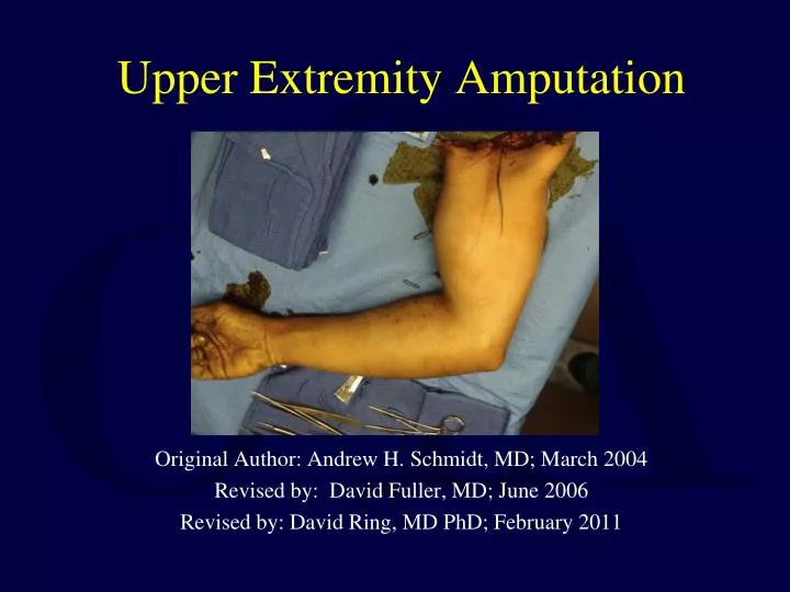
Upper Extremity Amputation Original Author: Andrew H. Schmidt, MD; March 2004 Revised by: David Fuller, MD; June 2006 Revised by: David Ring, MD PhD; February 2011
Amputation: Presentation Goals • Etiology • Techniques • Prosthetics and Rehabilitation
Amputation: Etiology • Trauma • Burns • Peripheral Vascular Disease • Malignant Tumors • Neurologic Conditions • Infections • Congenital Deformities
Etiology: Trauma • 90% of Upper Extremity Amputation • Male:Female = 4:1 • Most Amputations at level of Digit • Major Limb Amputations less common • Revascularization sometimes possible for incomplete amputation • Replantation sometimes possible for complete amputation
Etiology: Gangrene/Necrotizing Fasciitis Radiograph: Subcutaneous air throughout arm
Etiology: Vascular Disease Ischemia after AV Fistula Procedure
Etiology: Congenital polydactyly
Amputation: Trauma and Replantation • Candidates for Replantation after Trauma • 1. Thumb • 2. Multiple Digits • 3. Partial Hand • 4. Wrist or Forearm • 5. Above Elbow • 6. Isolated Digit Distal to FDS insertion • 7. Almost any part in child
Amputation: Trauma and Replantation • Candidates for Replantation after Trauma • Clean cut • Limited crush • Limited contamination • Acceptable ischemia time • 6 hours with muscle • 24 hours with digit
Surgical Technique: Digit Replantation • 1. Identify Vessels and Nerves • 2. Debride • 3. Shorten and fix bone • 4. Repair Extensor Tendon • 5. Repair Flexor Tendon • 6. Repair Arteries • 7. Repair Nerves • 8. Repair Veins • 9. Skin Closure (skin graft if necessary)
Amputation: Replantation • Poor Candidates for Replantation • 1. Severely crushed or mangled parts • 2. Multiple levels • 3. Other serious injuries or diseases • 4. Atherosclerotic vessels • 5. Mentally unstable • 6. > 6 hours ischemic time • 7. Severe contamination
Amputation: Replantation Mangled and Crushed – Poor Candidate
Ectopic “banking” of amputated parts • Indicated for extensive injuries with adequate amputated part in setting of contaminated or absent support structures. • Recipient sites described- anterior thorax, contralateral arm/leg, groin. High complication rate. • Largest and original series described by Marko Godina 1986.
Grip strength 80 # (unaffected side 100#) Injured right hand has remained dominant hand
Surgical Technique: Major Limb Replantation • Myonecrosis is greater concern than in digit replant • Immediate shunting to obtain arterial inflow may be necessary • High Potassium levels (>6.5 mmol/l ) in venous outflow from amputated part negative prognostic factor • Sequence of repair similar to digit • Identify structures, Debride, Rapid bone stabilization, Vascular repair (artery then veins), Tendons and Nerves
Upper vs Lower Limb • Upper extremity nonweightbearing • Less durable skin acceptable • Decreased sensation better tolerated • Joint deformity better tolerated • Late amputations rare • Transplants now being performed
Major Limb Replantation Include Surgical Prep of Legs for vascular and nerve grafts Rapid Bone Stabilization Ready for Anastomosis
UE traumatic amputation may be associated with life threatening hemorrhage Courtesy of T. Higgins, M. Dietch
Aggressive resuscitation and limb repair Courtesy of T. Higgins, M. Dietch
Amputation: Major Limb Replantation Outcomes • >2/3 survival rate • Can be a life threatening undertaking • Multiple Surgeries often required • Late Nerve, Bone, Tendon Surgeries • Function of major upper extremity replantations even though poor can be superior to prosthetic function
Outcomes: Major Limb Replantation • Comparison of functional results of replantation versus prosthesis in a patient with bilateral arm amputation Peacock, Tsai, CORR, 1987 • Major amputation of the UE: Functional Results after replantation/revascularization in 47 cases Daoutix et al, Acta Orthop Scand, 1995 • Major Replantation versus revision amputation and prosthetic fitting in the upper extremity: a late functional outcome study Graham et al, J Hand Surg, 1998
Amputation: Technique • Preservation of functional residual limb length balanced with • Soft tissue reconstruction to provide a well-healed, nontender, physiologic residual limb
Technique: Determination of Level • Zone of Injury (trauma) • Adequate margins (tumor) • Adequate circulation (vascular disease) • Soft tissue envelope • Bone and joint condition • Control of infection • Nutritional status
Tumor Forequarter Amputation
Necrotizing Fasciitis Emergent Open Shoulder Disarticulation
Trauma High Transhumeral Nerves Avulsed from High in Plexus
Failed Vascular Repair Transradial
Levels of Amputation • Wrist Disarticulation vs. Transradial • Disarticulation offers potential of better active pronation and suppination of forearm • Transradial often difficult to transmit rotation through prosthesis • Disarticulation poor aesthetically • Disarticulation more difficult to fit prosthetic • Transradial needs to be done 2 cm or more proximal to joint to allow prosthetic fitting • Transradial usually favored
Levels of Amputation • Transhumeral vs. Elbow Disarticulation • Adults: Elbow disarticulation allows enhanced suspension and rotation control of prosthesis however retention of full length precludes use of prosthetic elbow. Long transhumeral favored • Pediatrics: Transhumeral amputation results in high incidence of bony overgrowth. Elbow disarticulation is level of choice. Humeral growth slowed after trauma.
Levels of Amputation • Preservation of Elbow function is a priority • Consider replantation/salvage of parts to maintain elbow function • 4-5 cm of proximal ulna necessary for elbow function • For very proximal amputations, it may be necessary to attach bicep tendon to ulna
Techniques • Debridement of all Nonviable tissue and foreign material • Several debridements may be required • Primary wound closure often contraindicated • High voltage, electrical burn injuries require careful evaluation because necrosis of deep muscle may be present while superficial muscles can remain viable
Techniques • Nerve: Prevent neuroma formation • Draw nerve distally, section it, allow it to retract proximally • Skin: • Opportunistic flaps • Rotation flaps • Tension free • Skin grafts
Techniques • Bone: • Choose appropriate level • Smooth edges of bone • Narrow metaphyseal flare for some disarticulations Postoperative Dressing: • Soft • Rigid
Techniques • Goals of Postoperative Management • Prompt, uncomplicated wound healing • Control of edema • Control of Postoperative pain • Prevention of joint contractures • Rapid rehabilitation
Technique: Example 30 yo male, assault
Technique: Example ray amputation Be sure to identify all injuries and treat
Technique: Example 1 year postop
Technique: Example debridement and preservation of viable structure