Studying Aortic Stenosis: Impact on LV Remodeling
Investigating LV remodeling in aortic stenosis patients post-AVR. Explore arterial wave reflections and myocardial fibrosis correlation with MRI techniques. Testing hypothesis related to LV hypertrophy regression and physical fitness assessment.

Studying Aortic Stenosis: Impact on LV Remodeling
E N D
Presentation Transcript
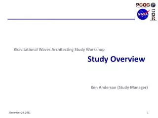
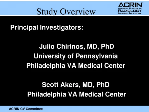
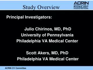
Study Overview Principal Investigators: Julio Chirinos, MD, PhD University of Pennsylvania Philadelphia VA Medical Center Scott Akers, MD, PhD Philadelphia VA Medical Center
Background • Calcific aortic stenosis is a disease of the elderly, which occurs in conjunction with age-related arterial stiffening and a consequent increase in pulsatile LV afterload. • Recently, it has been shown that the severity of aortic stenosis is not correlated with the degree of LVH assessed with MRI in patients with aortic stenosis,indicating that additional factors play an important role in this regard. • Regression of maldaptive LV remodeling post-AVR is variable Dweck MR. J Cardiovasc Magn Reson. 2012;14:50
LV Afterload (“Pressure overload”) • Important cause for LVH and LV failure. • In the absence of aortic stenosis, LV afterload depends entirely on the properties of the arterial tree. Gardin JM et al. Hypertension. 1997;29(5):1095-1103. Chirinos JA, Segers P. Hypertension 2010; 56(4):555-62.
Moens–Korteweg Equation Bramwell Hill Equation
A D1 D2 B
LV Afterload (“Pressure overload”) • Important cause for LVH and LV failure. • In the absence of aortic stenosis, LV afterload depends entirely on the properties of the arterial tree. Gardin JM et al. Hypertension. 1997;29(5):1095-1103. Chirinos JA, Segers P. Hypertension 2010; 56(4):555-62.
Afterload = Blood Pressure Vs. Blood Flow
Afterload = Blood Pressure Vs. Blood Flow • Patients with identical BP can have very different afterload patterns.
Afterload = Blood Pressure Vs. Blood Flow • Complex • “Steady” and Pulsatile components • Time-varying (time-resolved pressure and flow)
Aortic flow measurements Chirinos JA, Segers P. Hypertension 2010; 56(4):555-62.
Arterial determinants of the Central Pressure Profile Reflection timing Zc RM TAC 300 Flow (mm3/sec ) Pressure (mmHg) Reflection timing 240 180 SVR Zc 120 TAC 60 Reflection Magnitude 0 SVR Chirinos JA, Segers P. Hypertension 2010; 56(4):555-62. Chirinos JA, Segers P. Hypertension. 2010 56(4):563-70.
Effect of Early versus Late Pressure Overload in Rats A B Kobayashi S. Circulation. 1996;94:3362-3368.
0.035 Log-rank χ2 = 19.24 P < 0.0001 0.030 0.025 Top tertile Middle tertile Bottom tertile 0.020 Cumulative Hazard for First Heart Failure Event 0.015 0.010 0.005 0 0 2 4 6 8 Time to Heart Failure or Last Follow-up (Years) Chirinos JA et al. J Am CollCardiol2012.
0.030 0.025 HTN / High RM No HTN / High RM 0.020 HTN / Low RM No HTN / Low RM Cumulative Hazard for First Heart Failure Event 0.015 0.010 0.005 0 0 2 4 6 8 Time to Heart Failure or Last Follow-up (Years) Chirinos JA et al. J Am CollCardiol2012.
Predictors of incident heart failure in multivariate analysis (n=5932) Chirinos JA, Kips J, Jacobs D et al. JACC 2012.
Pusatile load and wave reflections cause maladaptive LV remodeling and hypertrophy and represent an important novel predictor of HF risk in the general population.Is it important in the setting of AS?
Primary Aim • To test the hypothesis that increased stiffness of the aortic wall and arterial wave reflections correlate with an adequate regression (improvement) of LV hypertrophy and LV myocardial fibrosis measured with cardiac MRI after AVR for severe aortic stenosis.
Secondary Aims • To test the hypothesis that myocardial T1rho mapping, a non-contrast myocardial tissue characterization MRI technique, correlates with LV myocardial fibrosis assessed with post-gadolinium myocardial T1 measurements in participants with severe aortic stenosis. • To test the hypothesis that changes in myocardial T1rho after AVR in participants with severe aortic stenosis correlates with changes in LV myocardial fibrosis assessed with post-gadolinium myocardial T1 measurements. • To test the hypothesis that increased stiffness of the aortic wall and arterial wave reflections correlate with physical fitness (assessed via a 6-minute walk test) after AVR for severe aortic stenosis
Inclusion Criteria • 18 years of age or older; • Severe symptomatic aortic stenosis (estimated aortic valve area <1 cm2) 1,2 • Planned for AVR between 0 and 28 days after enrollment; • Able to have a cardiac MRI between 7 and 28 days after enrollment (the 21 days before the AVR operation); • A preoperative coronary angiography demonstrating the absence of hemodynamically-significant CAD in need of revascularization during AVR; • Able to tolerate cardiac MR imaging with gadolinium contrast as required by protocol, to be performed at an ACRIN-qualified facility and scanner; • Willing and able to provide a written informed consent.
Exclusion Criteria • Not suitable to undergo cardiac MRI or gadolinium contrast administration (Claustrophobia, presence of metallic objects or implanted medical devices, etc) • Known allergy to contrast media • Weight greater than that allowable by the MRI table; • LV ejection fraction <50%; • Previous aortic valve surgery • Planned additional (non-aortic) valve repair/replacement • Infective endocarditis; • > Mild Aortic Insufficiency • Rhythm other than sinus rhythm • Presence of hemodynamically-significant CAD • Myocardial infarction or unstable angina in the previous month;
Exclusion Criteria • Pre-operative estimated glomerular filtration rate (eGFR) <40 mL/min/1.73m2 of body surface area. • An eGFR < 30 mL/min/1.73m2 of body surface area or acute kidney injury willl be a contrainidcation for gadolinium contrast administration at any time during the study. • Presence of a bicuspid aortic valve; • Resting heart rate >120 beats per minute, systolic blood pressure >180 mm Hg, or diastolic blood pressure > 100 mm Hg; • Pregnancy. • Unwillingness to undergo a cardiac MRI; • Unwillingness to sign the consent form. • Life expectancy < 1 year
VISIT 1: Eligibility/Registration • ICF • Eligibility • Medical History • eGFR within last 4 weeks • Review results of preop coronary angiogram • Exclude pregnancy • ECG within 8 weeks (NSR) • Data collection • KCCQ • Resgistration and Scheduling of baseline MRI
VISIT 2: Pre-AVR MRI • Recheck eGFR unless available within 4 weeks • Exclude pregnancy as per instutional standard of care • No smoking, ETOH or food for 4 hours or caffeine for at least 24 hours. • No nitrates for at least 4 hours • Usual medications • Blood draw (3 tubes: plasma, serum, htc). Store serum and plasma samples at -80 C in 0.5 mL vials.
VISIT 2: Pre-AVR MRI • IV line for Gad • Perform Cardiac MRI • Perform arterial tonometry • Phone interview 72 hours after visit for AEs
VISIT 3: 6 months post-AVR • KCCQ • eGFR check within 4 weeks (may require blood draw specifically for this purpose) • Exclude pregnancy • No smoking, ETOH or food for 4 hours or caffeine for at least 24 hours. • No nitrates for at least 4 hours • Usual medications • Blood draw (3 tubes: plasma, serum, htc). Store serum and plasma samples at -80 C in 0.5 mL vials.
VISIT 3: 6 months post-AVR • IV line for Gad • Perform Cardiac MRI • Perform arterial tonometry • 6-minute walk test • Phone interview 72 hours after visit for AEs
Arterial Tonometry Preston Broderick, MA University of Pennsylvania
Arterial Tonometry • Non-invasive device used to obtain Pulse Wave Analysis (PWA), Pulse Wave Velocity (PWV) • To be performed immediately after the MRI • ~30 minute procedure • Participant must be laying down • Do not include PHI in the database
Arterial Tonometry Adding a subject to the database
Arterial Tonometry • Information that must be entered • ID • First Initial • Last Initial • Year of Birth • Sex
Arterial Tonometry • Pulse Wave Analysis • Obtain BP measurements using an Omron HEM-907 device • Use that data to calculate the mean arterial pressure with the following equation: • MAP = DBP + 0.4 (SBP-DBP) • Type the sbp in the notes section as “SBP: x” • Indicate which side the radial tonometry is being performed in the notes section • Enter data into PWA subject screen as follows
Arterial Tonometry Pulse Wave Analysis (Radial)
Arterial Tonometry • Data acquisition • Relax your arms and hands • Maintain a constant pressure • Acquire 12 seconds of data • Complete acquisition by pressing the space bar • Goal is to achieve good consistency and height in the waveforms • Shoot for as high an operating index as possible • Ideal target: >90 for radial • Ideal target: >80 for carotid
Arterial Tonometry 12 Seconds of consistent data acquisition
Arterial Tonometry Favorable Result: 94 Operator Index
Arterial Tonometry Unfavorable Result: 0 Operator Index
Arterial Tonometry Pulse Wave Analysis (Carotid)
Arterial Tonometry Pulse Wave Analysis (Carotid)