Download
1 / 33
490 likes | 985 Views
Bone Health and Assessment in Children. Virginia Stallings, MD The Children’s Hospital of Philadelphia University of Pennsylvania School of Medicine. Overview. Assessing bone health in children: Quantitative Ultrasound (QUS) Dual Energy Xray Absorptometry (DXA)

E N D
Bone Health and Assessment in Children Virginia Stallings, MD The Children’s Hospital of Philadelphia University of Pennsylvania School of Medicine
Overview • Assessing bone health in children: • Quantitative Ultrasound (QUS) • Dual Energy Xray Absorptometry (DXA) • Peripheral Quantitative Computed Tomography (pQCT) • DXA – Distal Femur site • Body Composition • Development of a Calcium Food Frequency Questionnaire • Vitamin D status
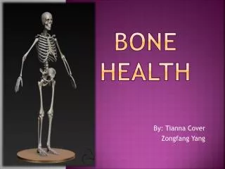
Bone Health Assessment Children are not little adults! http://www.bowhouse.com.au
Tibia – Sunlight Omnisense Phalanges – IGEA Calcaneus – Sahara, Cuba Ultrasound Sites and Techniques From Baroncelli 2008, Pediatric Research 62(3): 220-228.
Principles of QUS • Properties of the ultrasound wave (shape, amplitude) are altered by the material through which it passes in a way that is characteristic of the structural properties of the material • Material density and biomechanical properties (elastic modulus, compressive strength) of bone influence QUS waves
Principles of QUS • Trabecular bone scatters energy of ultrasound waves • Cortical bone absorbs energy of ultrasound waves
Principles of QUS • QUS applied to peripheral sites (calcaneus, phalanges, patella, radius, tibia) • Most ultrasound devices use a transmitter and detector to measure attenuation of the ultrasound wave • Critical angle reflectometry uses a single probe to measure the reflected wave as it travels along cortical bone and determine the speed of sound
Speed of Sound SoS – speed of sound; AD-SoS – amplitude dependent SoS; BTT – Bone transmission time (independent of soft tissue) From Baroncelli 2008, Pediatric Research 62(3): 220-228.
Sunlight Omnisense • Portable • Versatile – can measure radius or tibia • Probe sizes for different ages/sizes • Pediatric reference data from Israel
QUS Pediatric Reference Data Tibia Radius From Zadik et al. Osteoporosis International (10):857-62, 2003
Quantitative Ultrasound (QUS) • Advantages: • Rapid assessment • Radiation-free • Inexpensive and portable • Easily accessible measurement sites • Can be used in subjects without sleep/sedation • Limitations: • What are the bone properties (cortical vs trabecular) being measured? • Is it stable within an individual, and predictive of fracture?
Dual energy x-ray absorptiometry - DXA • Bone Mass, gm • Bone area, cm2 • Bone Density, gm/cm2
Advantages of DXA • Non-invasive test for measurement of BMD • Rapid, safe, easily tolerated, very low radiation exposure • Can assess bone density at different skeletal sites, both axial and peripheral • Widely available (100,000 in operation) • Excellent precision in children and adults
Pediatric Applications for DXA • Hologic infant software - Whole Body Scan • Requires no movement (sleep or sedation) • Clothing / swaddling material can affect body composition results • No reference data for current generation of DXA scanners
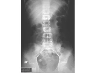
Pediatric Applications for DXA • Infant Spine scans • Not traditionally used for infants • Rapid scan time • Doesn’t require sedation • No body composition data, but clothing less of an issue
Why do both DXA and QUS • Tibia QUS is a measure of cortical bone • DXA spine scan is an integrated measure of cortical and trabecular bone • Studies of cortical and trabecular bone density changes during puberty stage suggest different effects in the 2 bone compartments
Why do both DXA and QUS • DXA can also be used for non-traditional measurement sites like the tibia to validate tibia QUS measurements • DXA is widely available and used clinically for children and adults, so study results may have greater clinical utility
Puberty Effects on Tibia Trabecular and Cortical Bone Density in Girls and Boys
Other Measurement Sites Distal Femur Distal Femur Radius
Cortical Density Cortical Area Bone Strength Bone Strength Index = (Moment of Inertia x Density) Peripheral Quantitative CT Total Density Total Area Cortical Density Cortical Area Trabecular Density Trabecular Area
66% 38% 3% Peripheral Quantitative CT Muscle Cross Sectional Area Cortical vBMD, dimensions and strength Trabecular & Total vBMD
Distal Femur DXA Scan • Indicated for: • Children with contractures • Indwelling hardware that would interfere with a spine or total body scan • Anomalies that would interfere with scan analysis or interpretation at standard sites • Might be especially useful for immobilized children who may be at increased risk of femur fractures Zemel et al J Clin Dens 2009 (in press) Henderson et al AJR Am J Roentgenol ;178:439
Development of the Calcium Counts Questionnaire • Conducted by interview with the assistance of food models and probing by interviewer • Recall over the previous 4 weeks because of the high intra-individual variability in calcium intake • Quantitative FFQ based on serving size and frequency of intake • First generation questionnaire: • Modified an existing calcium food-frequency questionnaire to capture foods that children usually eat based on recommendations of experienced pediatric research dietitians Zemel et al (in review)
Development of the Calcium Counts Questionnaire • Second generation questionnaire • Reviewed results of several hundred first generation questionnaires • Dropped items that were never selected • Reviewed the CSFII data for African Americans and Caucasians children – identified the top 50 sources of dietary calcium and added them to the questionnaire • Panel of pediatric research dietitians reviewed several nutrient content sources and assigned calcium values to items by consensus Zemel et al (in review)
Validation of the Calcium Counts Questionnaire • Compared results of the CCFFQ to 7-d weighed food diaries in 139 children 7 to 10 years of age • Conducted a test-retest over a 1 month interval to determine reproducibility • Concurrent validity was moderate (r=0.61) and test-rest reliability was high (r=0.74) • CCFFQ, like most FFQs, overestimated calcium intake compared to weighed food record Zemel et al (in review)
Gender differences in body composition From Butte et al. 2000 Ped Research 47(5):578-85
Pea Pod Infant Body Composition • Uses air displacement to determine body volume • With an accurate weight and volume measurement →body density • If assume that fat tissue has a constant density, →body composition derived from body density → fat-free mass, fat mass and percent body fat
Pea Pod Infant Body Composition • Rapid assessment • Safe, reliable, accurate, provides immediate results • Used up to measure infants up to ~4 to 6 months of age (1 to 8kg 1kg) • Length is the usual limtation
SUMMARY • Major advances in technology support bone health assessment in all ages • Optimal reference data are limited • Interpretation of results are complex in children with illness • Stature, FFM, velocity • Not just age and gender