Download
1 / 49
500 likes | 876 Views
OCULAR MANIFESTATIONS OF SARCOIDOSIS. Dr.Rajesh Babu B MS, FMRF, MSc (CEH) , DLSHTM, UK Consultant Uveitis & Ocular Immunology Ocular Epidemiology & Community Eye Health Narayana Nethralaya , Bangalore. Ocular Sarcoidosis. Definition Epidemiology Theories of pathogenesis

E N D
OCULAR MANIFESTATIONS OF SARCOIDOSIS Dr.Rajesh Babu B MS, FMRF, MSc (CEH) , DLSHTM, UK Consultant Uveitis & Ocular Immunology Ocular Epidemiology & Community Eye Health Narayana Nethralaya , Bangalore
Ocular Sarcoidosis • Definition • Epidemiology • Theories of pathogenesis • Clinical manifestations • Symptoms • Complications • Pathology • Differential diagnosis • Investigations • Treatment • Prognosis
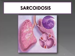
Definition • Sarcoidosis is a chronic multisystemic granulomatous disorder of unknown etiology thought to result from an exaggerated cellular immune response to a variety of self antigens or non-self antigens that is characterized by its pathological hallmark, the noncaseatinggranuloma.
The illness can be self-limited or chronicEpisodic recrudescence and remissions. • Because the lungs and thoracic lymph nodes are almost always involved, most patients report acute or insidious respiratory problems, variably accompanied by symptoms affecting the skin, eyes, or other organs.
Epidemiology • Sarcoidosis occurs worldwide. • Affecting persons of all races, both sexes, and all ages. • It has a particular proclivity for adults< age of 40 and for certain ethnic and racial groups (US blacks, Scandinavian and Irish white people). • The lifetime risk of sarcoidosis for U.S. blacks is 2.4 percent, and that for U.S. whites 0.85 percent.
Epidemiology • The frequency of sarcoidosis is reported to be low in various parts of the world. • It is not known whether this low frequency of sarcoidosis is genuine or whether it represents an underdiagnosis owing to the frequent occurrence of subclinical course, similarity with other diseases, or absence of firm diagnostic criteria.
Etiopathogenesis • The cause of sarcoidosis remains obscure for a number of reasons • The heterogeneity of the manifestations of the disease • The lack of a precise definition • Clinical overlap with other disorders • and insensitive and nonspecific diagnostic tests that lead to misclassification of the disease . • Mycobacterium remain leading suspects. • Transmission after cardiac /bone-marrow transplant has been reported • Almenoff PL, Johnson A, Lesser M, Mattman LH. Growth of acid fast L forms from the blood of patients with sarcoidosis. Thorax 1996;51:530-533. • Burke WMJ, Keogh A, Maloney PJ, Delprado W, Bryant DH, Spratt P. Transmission of sarcoidosis via cardiac transplantation. Lancet 1990;336:1579-1579. • Heyll A, Meckenstock G, Aul C, et al. Possible transmission of sarcoidosis via allogeneic bone marrow transplantation. Bone Marrow Transplant 1994;14:161-164.
Theories of pathogenesis The diverse manifestations of this disorder help fuel the prevailing hypothesis that sarcoidosis has more than one cause, each of which may promote a different pattern of illness.
Theories of pathogenesis Conceptually, it appears likely that Genetically predisposed hosts are exposed to antigens that trigger an exaggerated cellular immune response and the formation of granulomas.
PATHOLOGICAL AND IMMUNOLOGIC FEATURES Non-caseatinggranulomaholds the key to the diagnosis of sarcoidosis and provides clues to the immunopathogenesis of the disease.
Clinical Manifestations • Can be widespread or may involve only one organ system at a time. • Two peaks of incidence - the first at ages 20–30 years and the second at ages 50–60 years. • Ocular involvement manifests in 25%–60% of patients with systemic sarcoidosis. Albert DM, Jakobiec FAHunter DG, Foster CS(1994) Ocular manifestations of sarcoidosis. in Principles and practice of ophthalmology . eds Albert DM, Jakobiec FA(WB Saunders, Philadelphia), pp 443–450.
Symptoms • The majority of patients, however, present with systemic symptoms such as fatigue, anorexia, weight loss, and fever . • Many report dyspneaon exertion, retrosternal chest pain, and cough . • This may precede of come many months and years later. • Ocular symptoms related to chronic granulomatous uveitis • Lid nodules, conjunctival injection and nodules, rarely proptosis
Symptoms In 20 to 50 percent of the patients with more acute presentations, one sees the constellation of ErythemaNodosum Bilateral hilarlymphadenopathy Polyarthralgias, known as Löfgren's syndrome. Parotid involvement with uveitis is called Herfordt syndrome.
Examination findings • Ophthalmic lesions develop in approximately 25 percent of patients. • The classic symptom of anterioruveitis has a rapid onset, with blurred vision, photophobia, and excessive lacrimation; it usually clears spontaneously within a year. • Conjunctival involvement with small, pale, yellow nodules is common.
The most common ocular manifestations are uveitis (30%–70%) and conjunctival nodules (40%) • More than 80% of uveitis cases manifested before or within 1 year after the onset of systemic disease.
DIFFUSE MUTTON FAT KERATIC PRECIPITATES • AQUEOUS FLARE ++ • AQUEOUSCELLS ++ • FIBRIN PRESENT • IRIS NODULES • LENS CLEAR • NO VITREOUS REACTION RIGHT EYE
Conjunctival involvement has been reported in 6.9 - 70% of patients with ocular sarcoidosis Sarcoidosis granulomas are described as solitary, yellow "millet-seed" nodules. CONJUNCTIVAL NODULES
Posterior segment manifestations Intermediate Uveitis
Posterior segment manifestations Disc edema Active sarcoid granulomas
Complications • The clinical impact of sarcoidosis is directly related to the extent of granulomatous inflammation and its effect on the function of vital organs. Specimens should be obtained from the most readily accessible organ with the least invasive method.
Complications • The clinical impact of sarcoidosis is directly related to the extent of granulomatous inflammation and its effect on the function of vital organs. Specimens should be obtained from the most readily accessible organ with the least invasive method.
Pathology Small focus of necrosis. The most common appearance of necrosis in sarcoidosis
Rather like a rugby scrum with all the players grouped around the ball; only with sarcoidosis the ball is invisible.
Angiotensin converting enzyme (ACE) • ACE is normally present in the vascular endothelium of many organs (lung, kidney, small intestine, uterus, prostate, thyroid, testes, adrenals) and in macrophages. • The macrophages accounts for elevated ACE levels in patients with sarcoidosis. • ACE is elevated in 60-90% of patients with active sarcoidosis. • A normal serum ACE does not exclude the diagnosis, especially if the disease is in its early stages, localized to a small area (e.g. the eye), and therefore with a small epithelioid cell "burden".
False low values of ACE • Patients taking ACE inhibitors • Endothelial abnormalities, such as deep vein thrombosis • Patients who have had chemotherapy or radiation. • Treatment with systemic steroids or other immunosupressive agents can also affect ACE levels with values normalizing with adequate control of intraocular inflammation.
Elevated serum ACE • Gaucher's disease • Leprosy • Chronic pulmonary disease • Rheumatoid arthritis • Spondylitis • Primary biliary cirrhosis • Tuberculosis • Histoplasmosis • Histiocytic medullary fibrosis • Hyperthyroidism • Diabetes mellitus.
Differential diagnosis • Any cause of chronic granulomatous diseases • Tuberculosis • Syphillis • Herpetic uveitis • Lymphoma • Metastases
Investigations • In the absence of a known causative agent, sarcoidosis remains a diagnosis of exclusion. • There are no definitive diagnostic blood, skin, or radiologic imaging tests specific for the disorder. • Even serum ACE and galluium 67 Scan s are not specific to Sarcoidosis • Gold standard is demonstration of non caseatinggranulomas from tissue .
Investigations • CBC, ESR • Mantoux test • Chest X Ray • Serum ACE levels • Lymph Node Biopsy • Broncheo Alveolar Lavage • Trans Bronchial Lung Biopsy • Other tests • TPHA • Sputum for AFB • Gallium 67 Scan
Gold standard • In most centers, skin and transbronchial lung biopsies have supplanted biopsy of mediastinal lymph nodes and the liver because of their high yield, greater specificity, and low morbidity.
Lungs and thoracic lymph nodes are affected in 90% of patients with sarcoidosis Clinical staging is based on the pattern of chest radiographic findings: Radiographic Staging of Pulmonary Sarcoidosis Using Conventional Chest Radiography
Stage 1- Hilar and paratracheal adenopathy without parenchymal involvement. Bilateral hilar adenopathy is most common, followed by adenopathy in the right paratracheal area or aortopulmonary window area on the left.
Bilateral hilar adenopathy with Mild interstitial disease in upper lobes.
Stage 2-Adenopathy with parenchymal involvement. The parenchymal involvement includes a diffuse accentuation of interstitial markings resulting in a reticular pattern.
CT showing bilateral nodular infiltrates along bronchovascular bundles and enlarged mediastinal lymph nodes
Treatment • The optimal therapy for sarcoidosis is not well defined; therapeutic decisions are dictated by the localisation of the disease and severity of organ involvement. • The mainstay of treatment is corticosteroid therapy, which exhibits especially short term beneficial effects.
Treatment • Steroid sparing drugs for longer term treatment • Methotrexate 10-22.5mg/week • Cyclosporin B 3-5 mg/kgBW/day • Azathioprine 1-3mg/kgBW BID/TID
Local Treatment • Topical Prednisolone acetate eye drops • Peri-ocular Posterior subtenons triamcinolone acetonide • Dexamethasone implant in very severe recurrent cases
Prognosis • The poor visual prognosis was associated with the advanced age of the patients, black race, female sex, chronic systemic disease, and also with posterior segment involvement, peripheral punched out lesions, and the presence of cystoid macular oedema and glaucoma. • CME is leading cause of visual loss in sarcoid patients
Thank You drrajeshbabu@yahoo.com