Download
1 / 47
470 likes | 488 Views
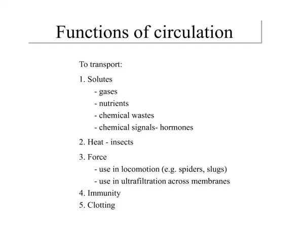
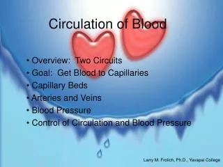
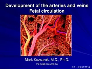
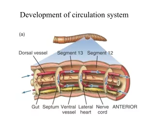
Development of circulation system. Heart. Ostium. Aorta. Hemocoel. Dorsal blood vessel. Gut cavity. Ventral blood vessels. Two of five hearts. Circulatory systems can be divided into two broad categories: open and closed.

E N D
Heart Ostium Aorta Hemocoel
Dorsal blood vessel Gut cavity Ventral blood vessels Two of five hearts
Circulatory systems can be divided into two broad categories: open and closed. Open circulatory system: blood pumped by the heart empties via an artery into an open, fluid-filled space, the hemocoel. Pressure are low. Some invertebrates Closed circulatory system: blood flows in a continuous circuit of tubes from arteries to veins through capillaries. All vertebrates and some invertebrates.
Plasma = 55% of whole blood Platelets “Buffy coat” <1% White blood cells Packed cell volume, or hematocrit Red blood cells = 45% of whole blood Fig. 9-3, p.361
Movement of blood results from any or all of the following mechanism • Forces imparted by rhythmic contractions of the heart (vertebrate) • Elastic recoil of arteries following filling by the action of heart (vertebrate) • Squeezing of blood vessels during body movement (arthropods) • Peristaltic contractions of smooth muscle surrounding blood vessels (giant earth worm).
Functions of the Circulatory System • Transportation: • Respiratory: • Transport 02 and C02. • Nutritive: • Carry absorbed digestion products to liver and to tissues. • Excretory: • Carry metabolic wastes to kidneys to be excreted in the urine.
Functions of the Circulatory System • Regulation: • Hormonal: • Carry hormones to target tissues to produce their effects. • Temperature: • Divert blood to cool or warm the body.
Functions of the Circulatory System • Protection: • Clotting: • Prevents blood loss. • Immune: • Leukocytes (antibodies and T cells), cytokines and complement fixation protect against pathogens.
Components of Circulatory System • Circulatory system consists of 4 basic parts: • A main propulsive organ, heart, force blood through body • An arterial system: distribute blood, pressure reservoir • Capillaries: transfer material between blood and tissues • A venous system: blood storage, returning blood to the heart
Pulmonary and Systemic Circulations • Pulmonary circulation: • -Path of blood from right ventricle through the lungs and back to the heart. • Systemic circulation: • Oxygen-rich blood pumped to all organ systems to supply nutrients.
desmosome Sarcoplasmic reticulum Gap junction Intercalated disc T-tube Carduac myofubril
Intrinsic Conduction System SA Node Internodal Pathway AV Node AV Bundle Bundle Branches Purkinje Fibers
Conduction of Impulse • AP from SA node spread quickly at rate of 0.8 - 1.0 m/sec. • Time delay occurs as impulses pass through AV node. • Slow conduction of 0.03 – 0.05 m/sec. • Impulse conduction increases as spread to Purkinje fibers at a velocity of 5.0 m/sec. • Ventricular contraction begins 0.1 – 0.2 sec. After contraction of the atria.
Electrocardiogram (ECG): A record of electrical events associated with contractions of the heart; typically obtained with electrodes placed on the surface of the body.
Electrical Activity of the Heart • Automaticity: automatic nature of the heartbeat. • SA node: • Demonstrates spontaneous depolarization. • Functions as the pacemaker. • Does not maintain a stable resting membrane potential. • Membrane depolarizes from –60 to –40 mV.
Depolarization • Depolarization: • VG fast Ca++ channels open. • Ca++ diffuses inward. • Opening of VG Na+ channels may also contribute to the upshoot phase of the AP. • Repolarization: • VG K+ channels open. • K+ diffuses outward.
Cardiac Muscle AP • Resting membrane potential of –90 mV. • SA node AP spreads to myocardial cells. • When myocardial cell reaches threshold, the cell depolarizes. • Rapid upshoot occurs: • VG Na+ channels open. • Inward diffusion of Na+.
Cardiac Muscle AP • Plateau phase: • Rapid reversal in membrane polarity to –15 mV. • VG Ca++ channels open. • Slow inward flow of Ca++ balances outflow of K+. • Rapid repolarization: • VG K+ channels open. • Rapid outward diffusion of K+.
A. Initiation of action potential in autorhythmic cells: 1. Pacemaker Potential due to influx of sodium and reduced efflux of potassium. 2. Depolarization and reversal of the membrane potential due to influx of calcium. 3. Repolarization due to efflux of potassium. B. .Initiation of action potential in contractile cells: 1. Opening of voltage-regulated fast sodium channels triggered by entry of positive ions from adjacent cell: Depolarization due to rapid influx of sodium 2. Plateau produced by calcium influx balancing potassium efflux. 3. Repolarization due to efflux of potassium.
Pacemaker Potential • - 40 mV is threshold for producing AP. • Spontaneous diffusion caused by diffusion of Na+ through slow Na++ channels. • Lack of a stable resting potential • Multiple array of channels • Parasympathetic stimulation decrease rate • Sympathetic stimulation increase rate
Refractory Periods • Heart contracts as on single unit. • Contraction lasts almost 300 msec. • Refractory periods last almost as long as contraction. • Summation cannot occur.
Summary • The intrinsic conduction system of the heart initiates depolarization impulses. • Action potentials spread throughout the heart, causing coordinated heart contraction. • An ECG wave tracing records the electrical activity of the heart.
Cardiac Cycle • Refers to the repeating pattern of contraction and relaxation of the heart. • Systole: • Phase of contraction. • Diastole: • Phase of relaxation.
Cardiac output Cardiac output: volume of blood pumped per unit time from a ventricle Stroke volume: volume of blood ejected from a ventricle by each beat of heart Exercise increase heart rates, not the stroke volume