Acute Inflammation
300 likes | 460 Views
Acute Inflammation. Clotting cascade. Inflammation is war. Coordinated response to eliminate the cause and consequence of injury or infection (noxious agent) Involves immune and vascular systems Acute inflammation is a stereotyped response to recent or ongoing injury

Acute Inflammation
E N D
Presentation Transcript
Inflammation is war • Coordinated response to eliminate the cause and consequence of injury or infection (noxious agent) • Involves immune and vascular systems • Acute inflammation is a stereotyped response to recent or ongoing injury • Chronic inflammation is a response to prolonged problems, orchestrated by T-helper lymphocytes
Inflammatory process • Immunity ¹ inflammation • Purpose is 3 d’s: • Destroy • Dilute • Dam-off • Desired result is healing • Regeneration (hyperplasia) • Fibrosis (scarring) • Collateral damage may occur
Classical description • Ancient Roman, Celsus, described inflammation • Calor—heat • Rubor—redness • Tumor—swelling • Dolor—pain • Hunter described inflammation as good in 1790s • Virchow added a fifth clinical sign in 1800s • Functio laesa—loss of function • Metchnikoff discovered phagocytes 1883 • Ehrlich discovered mast cells, granulocytes 1878 as well as complement in 1899 • Metchnikoff and Ehrlich shared Nobel Prize 1908
Inflammatory stimuli • Physical injury • Cutaneous laceration, osseous fracture, sunburn, toxin • Necrosis • Resolution of regions of dead cells • Infection • Innate response followed by adaptive response to microbial invader • Immunological errors • Allergies—response to environmental substances • Autoimmunity—response to self
Acute vascular changes • Transient arteriolar constriction (seconds) • Nerve reflex, endothelin • Like the immediate first step of hemostasis • Vasodilation of arterioles (until resolution) • Histamine, bradykinin mediate rapid response • Sustained by prostaglandins and NO • Hyperemia, erythema • Transudation increases blood viscosity, slows flow • Stasis and congestion in venules allows cells to contact endothelium • Vascular leakage (minutes to hours to days) • Histamine, bradykinin mediate rapid response • Sustained by C3a, C5a, PAF, leukotrienes • Exudation results in local edema
Increased vascular permeability • Contraction of vascular endothelium—rounding of cells and widening of intercellular spaces • Immediate, transient response (15-30 min) • Stimulated by histamine, bradykinin, substance P • venules of 20 – 60 um diameter respond • Delayed prolonged leakage (radiation burns) • begins after 2 – 12 h delay, lasts hours – days • stimulated by cytokines and apoptotsis of injured skin cells • venules and capillaries respond • Immediate, sustained response (days) • caused by direct damage to vasular endothelium • venules, capillaries, arterioles respond • ended by hemostasis, thrombosis, regeneration • Neutrophil-induced damage (days) • caused when neutrophils adhere and emigrate
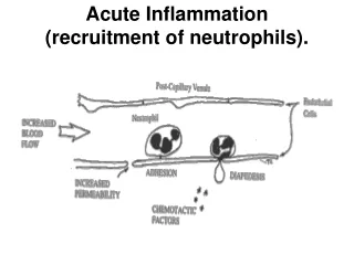
Leukocyte extravasation • Margination, rolling • Decreased flow rate and volume push WBCs toward vascular walls • Intermittent binding of selectins with glycoproteins causes rolling • Adhesion, pavementing • mediated by integrins on leukocytes, activated by cytokines • integrin ligands VCAM-1, ICAM-1 on endothelial cells induced by TNF and IL-1 • Transmigration or diapedesis • chemokines stimulate adherent leukocytes to migrate through interendothelial spaces
Selectins and Lewis X receptors • lectins are oligosaccharide binding proteins • selectins are a lectin-resembling class of glycoprotein receptors • selectin ligands contain an oligosaccharide called Lewis X and a sialic acid moiety
Regulation of expression of endothelial and leukocyte adhesion molecules A, Redistribution of P-selectin from intracellular stores to the cell surface. B, Increased surface expression of selectins and ligands for integrins upon cytokine activation of endothelium. C, Increased binding avidity of integrins induced by chemokines. Clustering of integrins contributes to their increased binding avidity (not shown). IL-1, interleukin-1; TNF, tumor necrosis factor.
Adhesion molecules • Integrins are ligands for CAMs • integrins are protein heterodimers • expressed on leukocytes • CD11/CD18,aka lymphocyte function-associated antigen 1 (LFA-1) binds ICAM • a4b1 binds VCAM • CAMs bind integrins • CAMs belong to the immunoglobulin superfamily • Intercellular CAM (ICAM) • Vascular CAM (VCAM) • Constitutively expressed by leukocytes • Made sticky by chemokines
Leukocyte transmigration • Extension of leukocyte pseupodia between endothelial cells • Binding to PECAM (CD31) expressed in endothelial junctions • Focal digestion of basement membrane with elastase, collagenase and metalloproteinases from neutrophils • Migration of leukocytes towards chemotactic gradient • Adhere to ECM with integrins and CD44
Chemotaxis of leukocytes • Locomotion oriented along a gradient of • Bacterial products • N-formyl peptides, lipoproteins, lipopolysaccharides • CXC chemokines • IL-8, aka CXCL8 or granulocyte chemotactic protein 1 • Complement proteins • Especially C5a • arachidonic acid (AA) metabolites • Leukotriene B4
Chemotactic receptors • Specific GPCRs • activation of second messengers • increase cytosolic calcium • activate Rac/Rho/cdc42 family GTPases • Result in movement • polymerization of actin • increased amounts of polymerized actin at the leading edge • localization of myosin filaments at the back
Which types of leukocytes respond • Neutrophils are generally the first types of leukocytes to respond in acute (non-viral) inflammation or to necrosis • Lymphocytes are usually the first cells to respond to viral infections or autoimmune diseases • lymphocytes and plasma cells also participate in most chronic inflammation, regardless of the inciting cause • Macrophages begin to appear a few days after the onset of inflammation from almost any cause, and increase in numbers over time • activated macrophages may develop abundant cytoplasm, called epitheloid macrophage • macrophages merge to create giant cells with multiple nuclei • Eosinophilic inflammation is highly suggestive of a response to helminths, arthropods, or allergens
Acute inflammation • Immediate, early response • Vasodilation • Vascular permeability • Emigration of leukocytes into tissues • Reactions of leukocytes in inflammation • Recognition of microbes and dead tissues • Removal of the offending agents • Macrophage activation • Leukocyte-mediated tissue injury
Receptors • Recognition of the offending agents • Toll-like receptors • lipopolysaccharide (LPS, or endotoxin), other bacterial proteoglycans and lipids, unmethylated CpG nucleotides • GPCRs • N-formyl peptides, complement (C5a), platelet activating factor, prostaglandins, and leukotrienes • Receptors for opsonins • IgG Fc, type 1 complement receptor (CR1) • Receptors for cytokines • IFN-γ is the major macrophage-activating cytokine • Activation the leukocytes • Increases in cytosolic Ca++ • Activation of protein kinase C and phospholipase A2
Activation of neutrophils • Stimulated by chemokines such as IL-8 and chemical mediators such as LTB4 • conformational change of integrin (LFA-1) to increase avidity for receptor (ICAM-1) • activation of Arachidonic Acid metabolic cascades • produces vasoactive prostaglandins and leukotrienes • activation of the oxidative burst to produce reactive oxygen species • NADPH oxidase enzyme system produces superoxide anion radical, which is converted to H2O2, which leads to production of hydroxyl radical • myeloperoxidase produces HOCl, which halogenates microbes • secretion of lysosomal enzymes (degranulation)
Actions of activated neutrophils • Phagocytosis has three steps • recognition and attachment of the particle to be ingested • engulfment, with subsequent formation of a phagocytic vacuole • killing or degradation of the ingested material • Initiation of repair • stimulate the proliferation of endothelial cells and fibroblasts • stimulate synthesis of collagen and enzymes that remodel connective tissues
Phagocytosis—Recognition • Mannose receptor • binds terminal mannose and fucose residues of glycoproteins and glycolipids (bacterial and fungal) • Scavenger receptors • oxidized or acetylated low-density lipoprotein (exogenous and endogenous lipid toxins) • High-affinity opsonin receptors • IgG antibodies—Fc receptor • C3b breakdown product of complement—C1R • mannan-binding lectin—C1R, CD14
Phagocytosis—Engulfment • Extensions of the cytoplasm (pseudopods) flow around bound receptors • Plasma membrane pinches off creating phagosome • Phagosome then fuses with a lysosomal granule • During this process the phagocyte may also release granule contents into the extracellular space • Engulfment is dependent on polymerization of actin filaments
Phagocytosis—Oxygen dependent killing • ROS generation is due to the rapid assembly and activation of NADPH oxidase (phagocyte oxidase), which reduces oxygen to superoxide anion • At least seven protein subunits located in the plasma membrane and the cytoplasm of resting neutrophils translocate to the phagosomal membrane to form the functional enzyme complex • Respiratory burst of G6PDH activity generates NADPH • Superoxide is converted to hydrogen peroxide (H2O2), mostly by spontaneous dismutation • Myeloperoxidase (MPO) from azurophilic granules converts H2O2 to hypochlorite (OCl•) with Cl- • NO generated by inducible nitric oxide synthase iNOS, aka NOS2 • NO reacts with superoxide to generate the highly reactive free radical peroxynitrite (ONOO•)
Phagocytosis—Oxygen independent killing • proteases, such as elastase and collagenase • defensins, cationic arginine-rich granule peptides that are toxic to microbes • lysozyme, which hydrolyzes the muramic acid–N-acetylglucosamine bond, found in the glycopeptide coat of bacteria • cathelicidins, antimicrobial proteins • lactoferrin, an iron-binding protein • major basic protein, a cationic protein of eosinophils, which is cytotoxic to many parasites • bactericidal/permeability increasing protein, which binds bacterial endotoxin
Damaging effects of phagocytes • Tissue damage occurs from • digestion of basement membranes during transmigration; caused by elastase and metalloproteinases • counteracted by a1-antitrypsin, C-reactive protein, and other circulating antiproteases • lysosomal leakage during phagocytosis (regurgitation or frustrated phagocytosis) • leaks acid hydrolases, free radicals, and hypochlorous acid • inflammatory mediators produced by phagocytes, such as prostaglandins and leukotrienes • necrosis of phagocytes in situ
Termination of acute response • Inflammation declines spontaneously • Mediators of inflammation are produced in rapid bursts only while the stimulus persists • Mediators have short half-lives and are degraded after their release. • Neutrophils also have short half-lives in tissues and die by apoptosis within a few hours after leaving the blood • Active termination mechanisms include • Switch from pro-inflammatory leukotrienes to anti-inflammatory lipoxins • Release of anti-inflammatory cytokines, including transforming growth factor-β (TGF-β) and IL-10, from macrophages • production of anti-inflammatory lipid mediators, called resolvins and protectins, derived from polyunsaturated fatty acids • neural impulses (cholinergic discharge) that inhibit the production of TNF in macrophages