Download

1 / 20
320 likes | 1.33k Views
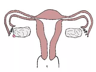
Pathology of the Fallopian tubes. Inflammations ( Salpingitis ) almost always bacterial in origin. Chlamydia , Mycoplasma , coliforms , (postpartum) strept . and staph. are now the major offenders.

E N D
Pathology of the Fallopian tubes • Inflammations (Salpingitis) • almost always bacterial in origin. • Chlamydia, Mycoplasma ,coliforms, (postpartum) strept. and staph. are now the major offenders. • Tuberculoussalpingitis is far less common and almost always with tuberculosis of the endometrium. • fever, lower abdominal or pelvic pain, and pelvic masses when the tubes become distended with either exudate or secretions. • Complications: • Adherence of tube to ovary tubo-ovarian abscess. • more seriousadhesions of the tubal plicae, and increasing the risk of tubal ectopic pregnancy. • Damage or obstruction of the tubal lumina may produce permanent sterility
Tubal malignancies • Currently, primary adenocarcinomas of the fallopian tubes are considered rare. • The most common is papillary serous carcinoma, others include endometrioid histology. • fallopian tube carcinomas are increased in women with BRCA mutations ( In studies of prophylactic oophorectomies:10% occult foci of malignancy usually in the fimbria). • Because the lumen and fimbria of the fallopian tube have access to the peritoneal cavity, fallopian tube carcinomas frequently involve the omentum and peritoneal cavity at time of presentation.
Ovarian Pathology • POLYCYSTICOVARIES (also called Stein-Leventhal syndrome). • oligomenorrhea, hirsutism, infertility, and obesity • usually in girls after menarche • secondary to excessive production of androgens by multiple cystic follicles in the ovaries (unclear causes). • Pathogenesis: excessive production of androgens; high concentrations of LH, and low concentrations of FSH. • The ovaries are enlarged, gray-white with a smooth outer cortex, and are studded with subcortical cysts 0.5 to 1.5 cm in diameter. • Histologically, there is a thickened, fibrotic outer surface, overlying cysts lined by granulosa cells with a hypertrophic and hyperplastic luteinized theca interna, with absence of corpora lutea.
Ovarian Neoplastic Diseases • Ovarian cancer is the fifth most common cancer in women. • It is also the fifth leading cause of cancer death in women. • Tumors of the ovary are diverse pathologic entities. • This diversity is attributable to the three cell types that make up the normal ovary: 1-The surface (coelomic) covering epithelium 2- The germ cells 3- The sex cord/stromal cells. • Each of these cell types gives rise to a variety of tumors
Pathogenesis-familial cases • Several risk factors for epithelial ovarian cancers have been recognized. Two of the most important are nulliparity and familyhistory. • prolonged use of oral contraceptives may reduce the risk. • Only 5%-10% of ovarian cancers are familial, the molecular pathogenesis of these cancers involve specific genes in these cases: • mutations in the BRCA genes 1 and 2. • The average lifetime risk for ovarian cancer approximates 30% in BRCA1 carriers, with figures varying from 16% to 44% in different studies. The risk in BRCA2 carriers is somewhat lower.
Pathogenesis- sporadic cases • Mutations in BRCA genes mutations are seen in only 8% to 10% of sporadic ovarian cancers. Thus, there must be other molecular pathways for ovarian neoplasms. • HER2/NEU is overexpressed in 35% of ovarian cancers (poor prognosis). • K-RAS protein is overexpressed in up to 30% of tumors, mostly mucinous cystadenocarcinomas. • p53 is mutated in about 50% of all ovarian cancers
SURFACE EPITHELIAL TUMORS • derived from the coelomicmesothelium that covers the surface of the ovary. • Benign lesions are usually cystic (cystadenoma) or can have an accompanying stromal component (cystadenofibroma). • Malignant tumors may be cystic (cystadenocarcinoma) or solid (carcinoma). • The surface epithelial tumors also have an intermediate, borderline category currently referred to as tumors of low malignant potential. These seem to be low-grade cancers with limited invasive potential. Thus, they have a better prognosis than the fully malignant ovarian carcinomas.
1- Serous Tumors • These are the most frequent ovarian tumors. • 60% benign, 15% borderline, and 25% malignant. • Benign serous lesions 30 and 40 years • malignant serous tumors 45 and 65 years of age. • Combined, borderline and malignant serous tumors are the most common malignant ovarian tumors and account for about 60% of all ovarian cancers. • Mutations in BRAF and K-RAS are common in borderline tumors and low grade cancers. • High-grade serous carcinomas mutations in p53 and BRCA1, and typically lack mutations in K-RAS and BRAF.
Morphology • Benign serous tumors: • large cystic structures, (30 to 40 cm in diameter). May be bilateral. The serosal covering is smooth and glistening. The cysts are usually filled with a clear serous fluid. characterized by a single layer of tall columnar epithelium. Some of the cells are ciliated. • Borderline serous tumors: • more complex architecture with mild cytologic atypiabutnostromal invasion. However, they might be associated with peritoneal implants. • Malignant serous carcinoma: Anaplasia of the lining cells and invasion of the stroma. • Note: Psammoma bodies (concentrically laminated calcified concretions) are common in the tips of papillae of serous tumors in general.
Prognosis of serous tumors: • Benign and borderline tumors have an excellent outcome (borderline tumors 100% survival, and even with peritoneal metastases it is nearly 75%, ). • Malignant invasive serous tumors prognosis is poor and depends heavily on the stage of the disease at the time of diagnosis.
2- Mucinous ovarian tumors • these tumors consists of mucin-secreting cells. • Only 10% of mucinous tumors are malignant (cystadenocarcinomas), while 10% are of low malignant potential (borderline), and 80% are benign. • Morphology • they are larger and multilocular. • psammoma bodies are not found • The cysts are lined by mucin secreting cells with abundant vaculated cytoplasm • Depending on the architectural complexity, these tumors are classified to benign, borederline or malignant. • The prognosis of mucinous cystadenocarcinoma is somewhat better than that for the serous counterpart, but the stage rather than the histologic type is the major determinant of treatment success.
3- Ovarian Endometrioid Carcinoma • Microscopically they are distinguished by the formation of tubular glands, similar to those of the endometrium, within the linings of cystic spaces. • Although benign and borderline forms exist, endometrioid tumors are usually malignant. • They are bilateral in about 30% of cases • 15% to 30% of women with these ovarian tumors have a concomitant endometrial carcinoma. • Similar to endometrial cancer, endometrioid carcinomas have mutations in the PTEN suppressor gene
Germ cell tumors • Benign (Mature) Cystic Teratomas: differentiation of totipotential germ cells into mature tissues representing all three germ cell layers: ectoderm, endoderm, and mesoderm. • Most are discovered in young women as ovarian masses or are found incidentally on abdominal scans because they contain foci of calcification produced by contained teeth. • 90% are unilateral • Grossly: often filled with sebaceous secretion and matted hair. Sometimes, foci of bone and cartilage, nests of bronchial or gastrointestinal epithelium, teeth and other recognizable lines of development are also present. • In 1% of cases there is malignant transformation of one of the tissue elements, usually taking the form of a squamous cell carcinoma. • these tumors are prone to undergo torsion (10% to 15% of cases), producing an acute surgical emergency
Clinical Correlations for All Ovarian Tumors • All ovarian neoplasms pose difficult clinical challenges, because they produce no symptoms or signs until they are well advanced. • The clinical presentation of all ovarian tumors is similar despite their great morphologic diversity • they cause local pressure symptoms (e.g., pain, gastrointestinal complaints, urinary frequency), sometimes they become twisted on their pedicles (torsion), producing severe abdominal pain mimicking an "acute abdomen." • Fibromas and malignant serous tumors often cause ascites,. • Functioning ovarian tumors often come to attention because of the hormones they induce.