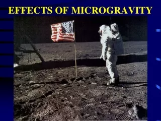
Impact of Microgravity on Fibroblast Cell Division: An Experimental Study
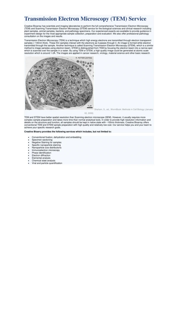
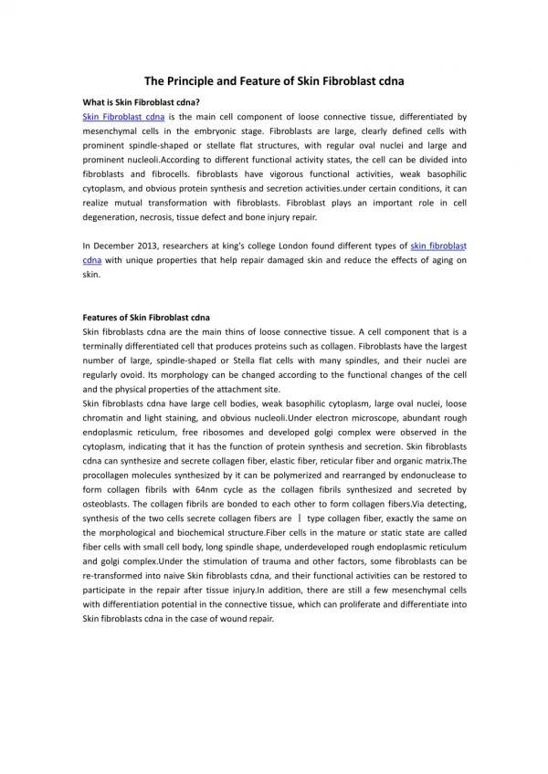
This study investigates how microgravity affects fibroblast cell division, a crucial biological process. Previous research indicates that simulated microgravity slows cellular division rates. Our hypothesis proposes that exposure to microgravity leads to decreased cell division due to microtubule disorganization, changes in actin filaments, less dense chromatin, and increased apoptosis. Using mouse embryonic fibroblasts cultured in specialized media, we conducted experiments to compare division rates in microgravity and normal gravity. Initial findings suggest that optimal cell growth occurs at 37°C with increased initial cell counts.

Impact of Microgravity on Fibroblast Cell Division: An Experimental Study
E N D
Presentation Transcript
fibroblast Division in Microgravity Jennifer Jiang, Jasmine Kuo, Kara Lukas San Marino High School San Marino, California
Background • Cell division is an integral component of life. • All organisms must go through the cell cycle in order to grow, develop, and reproduce. • Past experiments in simulated microgravity have shown that lack of gravity causes cells to divide at a slower rate.
Purpose • To discover the effect of microgravity on cell division • To compare the rates of cell division in the absence and presence of gravity
hypothesis • If cells are exposed to microgravity during their life cycles, then there will be a reduction in cell division because of • disorganization of microtubules • change in actin filaments • chromatin less dense • increased apoptosis
Materials • Type 3 FME • Mouse embryonic fibroblasts • Eagle’sMinimum Essential Medium (EMEM) • FetalBovine Serum (FBS) • Formaldehyde • Light Microscope • Hemocytometer • Trypan Blue
Assembly procedures Place experiment samples into the appropriate compartment of the FME. • Main Volume • 5.6 mL of EMEM • 0.56mL of 100% FBS • Short Ampoule A • 0.92 ml fibroblast cells • Short Ampoule B • 0.92 ml formaldehyde
experiment • Initiation: crack Ampoule A • Termination: crack Ampoule B • Experiment lasted 9 days
Data Number of fibroblasts
Analysis/Conclusion • Growth is more favorable for cells in 37°C CO2 incubator. • Cell growth is greater when the original cell count is increased from 500 to 750. • More conclusions will be drawn after flight experiment returns.
Acknowledgements • Mr. Wyeth Collo of San Marino High School • Dr. Susan Kane and Ms. Erin Denny of City of Hope Cancer Center • Center for the Advancement of Science in Space • San Marino Unified School District PTSA • Stephen and Mary Birch Foundation • Smithsonian Air and Space Museum • NanoRacks • Student Spaceflight Experiments Program