Download
1 / 48
520 likes | 689 Views
ANESTHETIC IMPLICATIONS OF HTN,CAD,ANAEMIA. Presented by- Dr. Kamal Prakash Sharma Moderator- Dr. Manoj Kumar Panwar. HYPERTENSION. Hypertension is most frequent preoperative abnormality in surgical patients, with an overall prevalence of 20–25%.

E N D
ANESTHETIC IMPLICATIONS OF HTN,CAD,ANAEMIA Presented by- Dr. KamalPrakash Sharma Moderator- Dr. Manoj Kumar Panwar
HYPERTENSION Hypertension is most frequent preoperative abnormality in surgical patients, with an overall prevalence of 20–25%. Definition: defined as two or more BP readings >140/90. Diagnosis of HTN cannot be made only by one preoperative reading but also requires confirmation by history of raised BP. BP also affected byposture, day time or night, emotional state, recent activity, drug intake, equipment and technique used. Thus preoperative anxiety, pain often produces some degree of hypertension even in normal patients but patients with a history of HTN have greater preoperative elevations in blood pressure.
Degree of end organ damage, morbidity and mortality correlates with duration and severity of HTN. Long standinguncontrolled hypertension accelerates atherosclerosis and hypertensive organ damage. Hypertension is a major risk factor for Cardiac, Cerebral, Renal, and Vascular, Ocular disease. Complications include Myocardial infarction, LVH, Congestive Heart failure, Stroke, Renal failure, Peripheral occlusive disease, and Aortic dissection. The presence of left ventricular hypertrophy (LVH) and carotid bruits—even in the absence of symptoms is an important predictor of cardiac mortality.
CLASSIFICATION OF BLOOD PRESSURE Category Systolic pressure Diastolic pressure Normal <130 <85 High normal 130-139 85-89 Hypertension Stage 1/Mild 140-159 90-99 Stage 2/Moderate 160-179 100-109 Stage 3/Severe 180-209 110-119 Stage 4/Very severe >210 >120
ETIOLOGY:- 1)Primary /Essential hypertension 2)Secondary hypertension Renal HTN Vascular Pyelonephritis, glomerulonephritis Diabetic nephropathy Endocrinal HTN Phaeochromocytoma Cushing’s disease Primary aldosteronism Vascular Coarctation of aorta Pregnancy induced hypertension
DRUG THERAPY Drug therapy has been shown to decrease progression of HTN, CAD,CHF and renal damage. Drug therapy also can reverse some pathophysiological changes like LVH and altered cerebral autoregulation. ORAL ANTIHYPERTENSIVE AGENTS 1)DIURETICS- Thiazide type Loop type Potassium sparing 2)SYMPATHOLYTICS – Adrenergic receptor blockers Central alfa-2 agonists Postganglionic blockers 3)VASODILATORS- Calcium channel blockers ACE inhibitors Angiotensin receptor blockers 4)DIRECT VASODILATORS- Hydralazine Minoxidil
Most patients with mild hypertension require only single-drug therapy, which may consist of a thiazide diuretic, ACE-inhibitor, ARB, beta-adrenergic blocker, or calcium channel blocker. The Joint National Committee on Hypertension recommends low doses of a thiazide diuretic for most patients. However, concomitant illnesses should influence drug selection. An ACE inhibitor is considered an optimal first-line choice for patients with left ventricular dysfunction or heart failure. ACE inhibitor or ARB is considered an optimal initial single agent in the setting of hyperlipidemia, chronic kidney disease or diabetes. Beta-blocker or less commonly, a calcium channel blocker is used as a first-line agent for patients with CAD.
ACE inhibitors, ARBs & adrenergic blockers are generally less effective than diuretics and calcium channel blockers in black patients. Patients with moderate to severe hypertension require a second or third drug. Preoperative Management It is generally recommended that elective surgery be delayed for severe HTN until BP is less than 180/110 mm Hg. If severe end organ damage is present, the goal should be to normalize BP as much as possible before surgery. Effective lowering of risk may require 6-8 weeks of therapy to allow regression of vascular and endothelial changes. If surgery can’t be postponed, the goal is not to decrease chronically increased BP too rapidly, as too rapid lowering of BP may increase risk of cerebral, coronary ischemia.
Thus decision to delay or to proceed with surgery should be individualized, based on the severity of HTN, coexisting myocardial ischemia, ventricular dysfunction, cerebrovascular or renal complications, surgical procedure (whether major surgically induced changes in cardiac preload or afterload are anticipated). History Severity and duration of the hypertension Drug therapy currently prescribed Compliance with the drug regimen. Presence or absence of hypertensive complications like myocardial ischemia, ventricular failure, impaired cerebral perfusion, or peripheral vascular disease. Adverse effects of current antihypertensive drug therapy
Physical Examination & Laboratory Evaluation BP measurement and auscultation Ophthalmoscopy Electrocardiogram Chest radiograph Echocardiography Renal function Serum electrolyte levels Premedication Premedication reduces preoperative anxiety and is highly desirable in hypertensive patients. Mild to moderate preoperative hypertension often resolves following administration of an anxiolytic agent, such as midazolam. Preoperative antihypertensive agents should be continued as close to schedule as possible and can be given with a small sip of water.
Intraoperative Management The overall anesthetic plan for a hypertensive patient is to maintain an appropriate stable blood pressure range. Blood pressure should generally be kept within 10–20% of preoperative levels. Patients with borderline hypertension may be treated as normotensive patients. Those with long-standing/poorly controlled HTN have altered autoregulation of cerebral blood flow, higher than normal MAP may be required to maintain adequate cerebral blood flow. Monitoring includes NIBP, ECG, SpO2, EtCO2, IBP, Urine output.
Induction Induction of anesthesia and intubation are often a period of hemodynamic instability for hypertensive patients. Regardless of the level of preoperative blood pressure control, patient’s with HTN display an accentuated hypotensive response to induction, followed by an exaggerated hypertensive response to laryngoscopy and intubation. Propofol, barbiturates, benzodiazepines, and etomidate are equally safe for induction in most hypertensive patients. Ketamine is contraindicated. Short duration laryngoscopy ≤15 seconds .
One of several techniques may be used before intubation to attenuate the hypertensive response: 1) Deepening anesthesia with a potent volatile agent for 5–10 min. 2) Administering a bolus of an opioid . 3) Administering lidocaine 1.5 mg/kg intravenously or intratracheally. 4) Achieving adrenergic blockade with esmolol 0.3–1.5 mg/kg, propranolol 1–3 mg or labetalol 5–20 mg. 5) Using topical airway anesthesia. Anesthesia may be safely continued with volatile agents (alone or with nitrous oxide), a balanced technique (opioid + nitrous oxide + muscle relaxant) With the possible exception of large boluses of pancuronium, any muscle relaxant can be used routinely. Hypertensive patients display an exaggerated response to both endogenous catecholamines (from intubation or surgical stimulation) and exogenously administered sympathetic agonists.
Intraoperative Hypertension Causes include light anaesthesia, hypoxemia & hypercapnia due to hypoventilation, CO2 insufflation during laproscopic procedures, excessive CO2 production. Cause of HTN should be sought and treated first before using antihypertensive agent Some parenteral agents for acute treatment of HTN Agent Dosage Range Onset Duration Nitroprusside 0.5-10 ug/kg/min 30-60 s 1-5 min Nitroglycerine 0.5-10 ug/kg/min 1 min 3-5 min Esmolol 0.5mg/kg over 1min f/b 50-300 ug/kg/min 1 min 12-20 min Labetalol 5-20 mg 1-2 min 4-8 h Propranolol 1-3 mg 1-2 min 4-8 h Nicardipine 0.25 -0.5 mg 1-5 min 3-4 h Nifedipine 10 mg 5-10 mg 4 hour Fenoldopam 0.1-1.6 mg/kg/min 5 min 5 min
Postoperative Management Postoperative hypertension is common and should be anticipated in patients who have poorly controlled hypertension. Close blood pressure monitoring should be continued in both the recovery room and the early postoperative period. In addition to myocardial ischemia and congestive heart failure, marked sustained elevations in blood pressure can contribute to the formation of wound hematomas and the disruption of vascular suture lines. Postoperative HTN may be due to pain, volume overload, hypoxemia, hypercapnia, bladder distention, hypothermia & shivering. Cause should be corrected and parenteral antihypertensive agents given if necessary. When the patient resumes oral intake, preoperative medications should be restarted.
CORONARY ARTERY DISEASE The overall incidence of CAD in surgical patients is estimated to be between 5% and 10%. Major risk factors for CAD Hyperlipidemia Hypertension Diabetes Cigarette smoking Increasing age Male sex Positive family history Obesity History of cerebrovascular or peripheral vascular disease Menopause Use of high-estrogen oral contraceptives Sedentary lifestyle CAD may be clinically manifested by symptoms of myocardial ischemia (usually angina), necrosis (infarction), arrhythmias(sudden death) or ventricular dysfunction (congestive heart failure).
Treatment of Ischemic Heart Disease The general approach in treating patients with ischemic heart disease is 5-fold: 1) Correction of coronary risk factors in the hope of slowing disease progression. 2) Modification of the patient's lifestyle to eliminate stress and improve exercise tolerance. 3) Correction of complicating medical conditions that can exacerbate ischemia, such as hypertension, anemia, hypoxemia, thyrotoxicosis, fever, infection, or adverse drug effects. 4) Pharmacological manipulation of the myocardial oxygen supply–demand relationship(nitrates, beta blockers, calcium channel blocker, aspirin) 5) Correction of coronary lesions by percutaneous coronary intervention or PCI (angioplasty with or without stenting, or atherectomy) or coronary artery bypass surgery.
PREOPERATIVE THERAPY:- OBJECTIVE To increase O2 supply to myocardium by maintaing diastolic BP Hb concentration maintaining O2 saturation To decrease determinants of myocardium O2 demand HR Contractility Ventricular wall tension Improved plaque stabilisation
DRUGS PRESCRIBED To continue beta blockers. Vasodilation with nitroglycerine, nitroprusside, prazosin in order to decrease ventricular wall tension. Allaying fear, anxiety and pain preoperatively are desirable goals in patients with CAD to prevents sympathetic activation, which affects myocardial oxygen supply–demand balance. A benzodiazepine alone or in combination with a opioid is commonly used. Concomitant administration of oxygen helps avoid hypoxemia following premedication. Preoperative Rx like statins, antihypertensives should generally be continued until the time of surgery.
ASPIRIN xcasdsdsdsds
INTRAOPERATIVE MANAGEMENT The basic challenges during induction and maintenance of anesthesia in patients with ischemic heart disease are (1) To prevent myocardial ischemia by optimizing myocardial oxygen supply and reducing myocardial oxygen demand. (2) To monitor for ischemia. (3) Totreat ischemia if it develops. Intraoperative Events That Influence Myocardial Oxygen supply-demand relationship Decreased Oxygen Delivery Decreased coronary blood flow Tachycardia Diastolic hypotension
Hypocapnia (coronary artery vasoconstriction) Coronary artery spasm Decreased oxygen content Anemia Arterial hypoxemia Shift of the oxyhemoglobin dissociation curve to the left Increased Oxygen Requirements Sympathetic nervous system stimulation Tachycardia Hypertension Increased myocardial contractility Increased afterload Increased preload
Choice of Anesthesia Regional Anesthesia Regional anesthesia is often a good choice for procedures involving the extremities, the perineum & possibly the lower abdomen. Precipitous decreases in blood pressure should be rapidly treated with small doses (25–50 ug) of phenylephrine or similar agent to preserve coronary perfusion pressure until sufficient intravenous fluid can be given. Marked hypotension can usually be avoided by prior volume loading. Small doses of ephedrine (5–10 mg) may be preferable in the presence of bradycardia.
Hypotension not responding to phenylephrine or ephedrine may be treated with small doses of epinephrine (2–10 ug). Patchy/incomplete surgical anesthesia/excessive sedation during regional anesthesia defeats the purpose of selecting a regional technique, unnecessarily stresses the patient & may precipitate myocardial ischemia General Anesthesia Induction should have minimal hemodynamic effects. produce reliable loss of consciousness. provide sufficient depth of anesthesia to prevent a vasopressor response to intubation.
Induction with small incremental doses of the selected agent usually avoids the precipitous decreases in blood pressure that can be seen following a large bolus. Titration of the induction agent—first against loss of consciousness and then to an acceptable decrease in blood pressure. Moreover, sufficient anesthetic depth for intubation can be achieved with less cardiovascular depression than that caused by the bolus technique. Administration of a muscle relaxant (as soon as the eyelid reflex is lost) & controlled ventilation ensure generally adequate oxygenation throughout induction. Endotracheal intubation is performed once sufficient anesthetic depth is reached or arterial blood pressure reaches its lowest acceptable limit.
Blood pressure, heart rate and the ECG should be repeatedly assessed with each step during induction. Induction Agents Propofol, barbiturates, etomidate, benzodiazepines, opioids, and various combinations of these drugs are often used. High-dose opioid anesthesia had previously been used widely for patients with significant ventricular dysfunction. Opioids used as sole agents may not be complete anesthetics because of high incidence of intraoperative awareness, hypertension, prolonged respiratory depression, chest wall rigidity following this technique & is unsuitable for most noncardiac operations.
Myocardial ischemia may accompany the sympathetic nervous system stimulation that results from direct laryngoscopy and tracheal intubation. Short-duration direct laryngoscopy (≤15 seconds) is useful in reducing the magnitude & duration of the circulatory changes associated with tracheal intubation. If the duration of direct laryngoscopy is not likely to be brief or if hypertension already exists, it is reasonable to consider administering drugs to minimize the pressor response. Maintenance of Anesthesia Tachycardia & HTN are likely to develop in response to intense stimulation, as during direct laryngoscopy or painful surgical stimulation.
Controlled myocardial depression using a volatile anesthetic may be useful in such patients to minimize the increase in sympathetic nervous system activity. The volatile anesthetic may be administered alone or in combination with nitrous oxide. Use of a nitrous oxide–opioid technique with the addition of a volatile anesthetic is equally acceptable to treat any increases in blood pressure that accompany painful surgical stimulation. Volatile anesthetics may be beneficial in patients with ischemic heart disease by virtue of decreasing myocardial oxygen requirements & preconditioning the myocardium to tolerate ischemic events detrimental because of decrease in BP and associated decreases in coronary perfusion pressure.
Patients with severely impaired left ventricular function may not tolerate anesthesia-induced myocardial depression. Opioids may be selected for these patients & addition of nitrous oxide, benzodiazepine or low-dose volatile anesthetic may be needed because total amnesia cannot be ensured with an opioid alone, but the addition of nitrous oxide/volatile anesthetic may be associated with myocardial depression. Choice of Muscle Relaxant The choice of muscle relaxant in patients with ischemic heart disease is also influenced by their impact on myocardial oxygen demand supply relationship.
Muscle relaxants with minimal or no effect on HR & BP (vecuronium, rocuronium, cisatracurium) are choices for patients with ischemic heart disease Histamine release and the resulting decrease in BP caused by atracurium are less desirable. Myocardial ischemia has been described in patients with ischemic heart disease given pancuronium, presumably because of increase in HR and BP produced by this drug. Reversal of muscle paralysis with standard agents does not appear to have any detrimental effects in patients with CAD. Use of glycopyrrolate decrease the likelihood of transient tachycardia
INTRAOPERATIVE MONITORING Perioperative monitoring is influenced by the complexity of the operative procedure and the severity of the ischemic heart disease. An important goal when selecting monitors is to select those that allow early detection of myocardial ischemia. Most myocardial ischemia occurs in the absence of hemodynamic alterations. 1) NIBP, SpO2, EtCO2 2) ELECTROCARDIOGRAPHY The diagnosis of myocardial ischemia focuses on changes in the ST segment characterized as depression or elevation of at least 1 mm.
T-wave inversion and R-wave changes can also be associated with myocardial ischemia, although other factors such as electrolyte changes can also produce such changes. The degree of ST-segment depression parallels severity of ischemia. Traditionally, monitoring two leads, II and V5, has been the standard, but it appears that monitoring three leads improves the ability to detect ischemia. Leads II, V4, and V5 or V3, V4, and V5 are the sets of three leads recommended. There is a predictable correlation between the lead of the ECG that detects myocardial ischemia and the anatomic distribution of the diseased coronary artery. 3) INTRAARTERIAL PRESSURE monitoring is advisable for all patients with severe CAD and with major or multiple cardiac risk factors.
4) CENTRAL VENOUS or PULMONARY ARTERY PRESSURE monitoring during prolonged or complicated procedures involving large fluid shifts/blood loss. Monitoring of pulmonary artery pressure may be desirable for patients with significant ventricular dysfunction (ejection fraction < 40–50%). 5) TRANSESOPHAGEAL ECHOCARDIOGRAPHY The development of new regional ventricular wall motion abnormalities is the accepted standard for the intraoperative diagnosis of myocardial ischemia. These regional wall motion abnormalities seen before ECG changes occur.
Intraoperative Management of Myocardial Ischemia Treatment of myocardial ischemia should be instituted when there are 1-mm ST-segment changes on the ECG. Prompt, aggressive pharmacologic treatment of changes in heart rate and blood pressure is indicated. A persistent increase in heart rate can be treated by intravenous administration of a β-blocker such as esmolol. Nitroglycerin is a more appropriate choice when myocardial ischemia is associated with a normal to modestly elevated blood pressure. In this situation, the nitroglycerin-induced coronary vasodilation and decrease in preload facilitate improved subendocardial blood flow but the nitroglycerin-induced decrease in afterload does not decrease systemic blood pressure to the point that coronary perfusion pressure is jeopardized.
Hypotension is treated with sympathomimetic drugs to restore coronary perfusion pressure. In addition to vasoconstrictor drugs, fluid infusion can be useful to help restore blood pressure. Regardless of the treatment, prompt restoration of blood pressure is necessary to maintain pressure-dependent flow through coronary arteries narrowed by atherosclerosis. In an unstable hemodynamic situation, circulatory support with inotropes may be necessary. It may also be necessary to plan for early postoperative cardiac catheterization.
Postoperative Management The goals of postoperative management are the same as those for intraoperative management: prevent ischemia, monitor for myocardial injury and treat myocardial ischemia/infarction. Intraoperative hypothermia may predispose to shivering on awakening, leading to abrupt and dramatic increases in myocardial oxygen requirements. Pain, hypoxemia, hypercarbia, sepsis & hemorrhage also lead to increased myocardial oxygen demand. The resulting oxygen supply/demand imbalance in patients with ischemic heart disease can precipitate myocardial ischemia, infarction or death. Although most adverse cardiac events occur within the first 48 hours postoperatively, delayed cardiac events (within the first 30 days) can also occur.
Prevention of hypovolemia and hypotension is necessary postoperatively & not only intravascular volume but also an adequate hemoglobin concentration must be maintained. Continuous ECG monitoring is useful for detecting postoperative myocardial ischemia, which is often silent. Postoperative myocardial ischemia should be identified, evaluated and managed aggressively, preferably in consultation with a cardiologist.
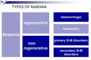
ANAEMIA Definition: A qualitative or quantitative decrease in the number of red blood cells (RBC's) or hemoglobin, resulting in a lower ability for the blood to carry oxygen to body tissues. In adults, anemia is usually defined as Hb concentrations less than 11.5 g/dL (hematocrit, 36%) for women and less than 12.5 g/dL(hematocrit, 40%) for men. CAUSES OF ANAEMIA A) Physiological anaemia of pregnancy B) Acquired: a. Nutritional- Iron deficiency, folate deficiency, B-12 deficiency, etc. b. Infections- Malaria, hookworm infestation, etc c. Haemorrhagic- Acute or chronic blood loss d. Bone marrow suppression- Aplasticanaemia,drugs, etc. e. Renal disease f. Haemolytic C) Genetic - haemoglobinopathies – sickle cell disease, thalassaemia, etc
PATHOPHYSIOLOGY OF ANAEMIA Oxygen is carried in the blood in two forms as: Physical solution in plasma (dissolved form).Arterial blood contains only 0.3 mL of O2, in each 100 mL of blood at a PO2 of 100 mm Hg and temperature of 37’C 2) Reversible chemical combination with haemoglobin/Oxyhaemoglobin Hb reversibly binds to four molecules of O2 which equals to 1.37-1.39 mL/g of Hb. O2 content of the blood is calculated from the equation: CaO2= Hb × 1.37 × SaO2 + 0.0034 × PaO2 mm Hg Total quantity of O2 in arterial blood delivered to tissues is a function of cardiac output (CO). Therefore, Oxygen delivery= CaO2 × Cardiac Index × 10mL/min/m2 Whenever anemia occurs, i.e., CaO2 decreases, CO increases as a compensatory mechanism to maintain O2 delivery to tissues.
O2 delivery is also affected by the relationship between the Hb saturation (SaO2) & the partial pressure of O2 in the blood, described as oxygen dissociation curve (ODC). Thus in an anemic pregnant patient various compensatory mechanisms get activated: 1. Increase in CO 2. Rightward shift of ODC 3. Decrease in blood viscosity 4. Increase in 2,3-DPG concentration in RBC 5. Release of renal erythropoietin leading to stimulation of erythroid precursors in bone marrow Thus, in physiological or chronic anaemia tissue oxygenation is not impaired as a result of compensatory mechanisms. May be compromised in severe or acute onset anaemia leading to serious consequences like right heart failure, angina, tissue hypoxemia.
ANAESTHETIC CONSIDERATIONS Preoperative assessment Clinical assessment should focus at assessment of the cause, type and severity of anemia and adequacy of compensatory mechanisms. History suggestive of poor tissue perfusion can manifest as tiredness, easy fatigability in mild anemia to SOB/dyspnoea, palpitations, angina in moderate to severe anemia. Signs of high CO like tachycardia, wide pulse pressure, systolic ejection murmur are essential for planning the mode of anaesthetic management. Investigations should include a complete haemogram, reticulocyte count, peripheral smears and blood grouping. Other investigations include ESR, blood urea nitrogen levels, S. creatinine, bilirubin levels, S. proteins, S. Iron, total iron-binding capacity, B12 and folate levels, Hb electrophoresis and ECG for any evidence of myocardial ischaemia, etc.
Minimal acceptable level of Hb and need for preoperative transfusion A ‘minimum acceptable haemoglobin level’ does not exist. A healthy myocardium compensates for the low Hb or Hct levels (7-8 gm/dL of Hb or 21-24% Hct)in order to optimize O2 delivery. In patients with overt or silent episodes of myocardial ischaemia, a level of < 10 gm/dL carries risk of decompensation. Many task force guidelines recommend that RBC transfusions should not be dictated by a single Hb trigger”; instead, it should be based on the patient’s needs and risks of developing complications of inadequate oxygenation. The American College of Surgeons recommends RBC transfusions to normovolemic patients with anemia only if symptoms are present.
An Hb level of 8 g/dL was suggested as a “transfusion trigger” by the Transfusion Practice Committee of the American Association of Blood Banks A threshold of 7 g/dL was suggested by the National Institutes of Health Consensus Conference on Perioperative Blood Transfusion. The decision to transfuse should be made on basis of symptoms, coexisting medical conditions, continuing blood loss or threat of bleeding Benefits from replenishing O2-carrying capacity by transfusion must always be balanced against transfusion-associated risks like pulmonary edema, immune suppression.
Choice of anaesthesia Depend on the severity, type of anaemia, extent of physiological compensation, concomitant medical conditions, type and nature of procedure and anticipated blood loss. The main anaesthetic considerations in chronic anaemia are to minimize factors interfering with O2 delivery, prevent any increase in O2 consumption and to optimize the partial pressure of O2 in the arterial blood. The following measures need to be diligently adhered to in the peri operative period A. Avoidance of hypoxia a. Preoxygenation is mandatory with 100% O2. b. Oxygen supplementation should be given in the perioperative period. c. Maintenance of airway is important to prevent fall in FiO2 due to airway obstruction, difficult intubation, etc. Hence measures and expertise to secure a definitive airway should be available immediately.
d. Spontaneous ventilation technique with High FiO2 (40-50%) to overcome effects of hypoventilationissuitableonly for short procedures. e. Aggressively treat and avoid conditions that increase the O2 demands like fever, shivering,acute massive bloodlosses. f. Nitrous oxide should be used cautiously in patients with folate and Vitamin B-12 deficiency. B. Minimize drug-induced decreases in CO a. Intravenous induction of anaesthesia should be slowly titrated to prevent precipitous fall in CO. b. Careful positioning of the patient to minimize position associated volume shifts. c. Mild tachycardia and wide pulse pressure may be physiological and should not be confused with light anaesthesia.
C. Factors leading to left shift of ODC should be avoided a. Avoid hyperventilation to minimize respiratory alkalosis. Hypocapnia also decreases CO. Maintain normocapnia. b. Hypothermia should be avoided – i. ensure normal core body temperatures ii. IV fluids and blood products if any should be warmed D. Monitoring:- aimed at assessing the adequacy of perfusion and oxygenation of vital organs. ECG, NIBP, EtCO2, Temperature monitoring, Pulse oximetry, urine output CVP, IBP monitoring, ABG analysis and measurement of mixed venous PvO2 in severe anemia where in major blood losses are anticipated. Serial Hb and Hematocrit values can guide us to monitor ongoing blood losses.
E. Regional anaesthesiais preferred for peripheral limb surgery as they are associated with reduced blood loss F. Central neuraxial blocks using either a low-dose spinal anaesthesia along with adjuvants or an intermittent dosing, continuous epidural. Advantages:- good analgesia ability to provide supplemental O2 decreased blood loss with stable haemodynamics. Disadvantages:- hypotension haemodilution and subsequent heart failure pulmonary edema on the return of vascular tone. It is advisable to use vasoconstrictors to sustain blood pressure. Regional anaesthesia can also be implicated in the worsening of symptoms of subacute degeneration of spinal cord and hence should be avoided inVitamin B12 deficiencies with neurological symptoms.