Download
1 / 67
700 likes | 799 Views
DNA - The Code of Life. Chapter 16. Bacterial Transformation. F. Griffith & Transformation. 1928 Streptococcus pneumoniae 2 strains: R – harmless, S – pathogenic Mixed inactive S strain & active R strain; injected into mouse Mouse died, pathogenic strain in blood Transformation.

E N D
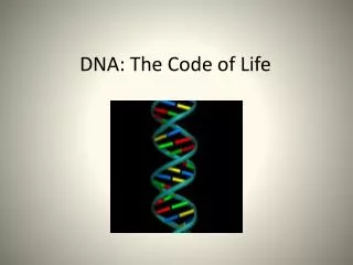
DNA - The Code of Life Chapter 16
F. Griffith & Transformation • 1928 • Streptococcus pneumoniae • 2 strains: R – harmless, S – pathogenic • Mixed inactive S strain & active R strain; injected into mouse • Mouse died, pathogenic strain in blood • Transformation
Avery, McCarty & MacLeod • 1944 : transforming agent is DNA • Skeptics – bacteria not complex • More research • Viruses (bacteriophages) • Viral structure & replication?
Hershey & Chase – The Blender Experiment • 1952: DNA is the genetic material of the phage T2 • T2 phage infects E. coli • Labeled protein coat with radioactive S and the DNA with radioactive P • Phages infect E. coli separately • Only P found in the bacterium
Findings • Infected with radio-labeled proteins - radioactivity in supernatant • Infected with radio-labeled DNA - radioactivity in pellet • Hershey & Chase’s conclusion?
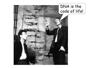
Let the race begin! • 1950s – scientific community racing to find 3-D structure of DNA • Major players • James Watson & Francis Crick • Linus Pauling • Maurice Wilkins & Rosalind Franklin
Puzzle Pieces • Chargaff’s Data • Backbone Structure – single strand • Franklin’s Data
Chargaff’s Rules - Structure • 1947 • Polymer - deoxyribose sugar, phosphate grp & nitrogen-containing base • Bases: • adenine (A), thymine (T), guanine (G), or cytosine (C)
Chargaff’s Rules • Certain bases were always equal in number • # adenines approx equal to # of thymines (%T = %A) • # guanines approx equal to # of cytosines (%G = %C) • Why is this significant?
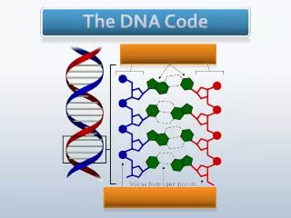
Backbone Structure • Phosphate group of one nucleotide attached to the sugar of the next • Result is a “backbone” of phosphates and sugars, from which the bases project
R. Franklin & M. Wilkins X-Ray Crystallography
Franklin’s Data • Wilkins & Franklin used X-ray crystallography to study DNA structure • X-rays diffracted as they pass through aligned fibers of purified DNA • Diffraction pattern used to deduce 3-D shape of molecules
What the picture tells us • DNA was helical in shape!
Putting the puzzle together… • Watson & Crick – “stolen” ideas or a product of the times? • However… it was Watson who deduced the width of the helixand the spacing of bases • Model building to beat Pauling
TRIAL AND ERROR • Watson & Crick began to work on a model of DNA with two strands, the double helix • Wire molecular models - first tried to place the sugar-phosphate chains on the inside • Did not fit the X-ray measurements and other info on chemistry of DNA
Breakthrough • Watson put sugar-phosphate chain on the outside & nitrogen bases on the inside
Nuts & Bolts – Chargaff’s Data • Watson & Crick determined that chemical side groups off nitrogen bases formed H bonds, connecting strands • Adenine - form 2 H bonds only with thymine • Guanine - form 3 H bonds only with cytosine • Findings explained Chargaff’s rules
DNA Structure • DNA bases: • Purines: adenine and guanine • Pyrimidines: thymine and cytosine • Nucleotides are covalently bonded with a sugar-phosphate backbone • The linkage forms a 3’,5’ phosphodiester linkage • One end of the molecule has a free 5’ carbon; the other has a free 3’ carbon
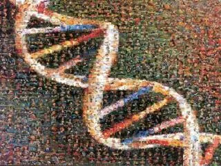
DNA Structure • Two polynucleotide chains intertwined to form a double helix
Technical DNA Data • .34 nm is the distance between the bases • 3.4 nm repeat of nucleotides due to a complete “turn” of the helix • width of molecule is 2.0 nm
Pyrimidines and Purines • Pyrimidines are single-ringed • Purines are double-ringed • Bonding is complementary • Sequence in one chain dictates sequence in opposite chain
DNA Replication • Complimentary bases act as templates • Bases of one strand allow for exact duplication
DNA Replication • Origin(s) of replication • Prokaryotes - single specific sequence of nucleotides recognized by replic. enzymes • Replication proceeds in both directions • Eukaryotes – hundreds/thousands of origin sites per chrom • Bubble with replication forks at each end • Bubbles elongate as DNA is replicated and eventually fuse
Bidirectional Synthesis • In prokaryotes, the circular DNA is opened up, and synthesis occurs in both directions
Bidirectional Synthesis • In eukaryotes, the linear DNA has many replication forks
DNA Replication • Proteins and enzymes work together • DNA strands must be unwound during replication • DNA helicase unwinds the strands • Single stranded binding proteins (SSB) prevent immediate reformation of the double helix • Topoisomerases break and then rejoin the strands, “untying” the knots that form
DNA Replication Order • Always proceeds in a 5’ 3’ direction • DNA polymerase can add only at the 3’ end • Nucleotides are polymerized and 2 phosphates are removed in the process
Nucleotide Synthesis • Raw nucleotides are nucleoside triphosphates • N base, deoxyribose, & a triphosphate tail • Nucleotide added, last 2 phosphate grps hydrolyzed, forming pyrophosphate • Exergonic rxn drives polymerization of the nucleotide to the new strand
DNA Pol • DNA polymerases can only add nucleotides to the free 3’ end of a growing DNA strand • A new DNA strand can only elongate in the 5’ 3’ direction
Problems? • One parental strand is oriented 3’ 5’ into the fork, while the other is oriented 5’ 3’ into the fork • At fork, only one parental strand (3’ 5’ into the fork), leading strand, can be used by polymerases as a template for a continuous complementary strand
Continuous & Discontinuous • Replication is continuous on one strand and discontinuous on the other • Replication begins at replication forks
Okazaki fragments • Synthesis of the leading strand is continuous • The lagging strand (discontinuous) is synthesized in pieces called Okazaki fragments
Okazaki fragments • 100 - 1000 nucleotides in length • Initiated by a separate RNA primer • Okazaki fragments are joined together by DNA ligase
RNA Primer • DNA pol cannot initiatesynthesis because it can only add nucleotides to end of an existing chain • Requires an RNA primer • Primase, an RNA pol, links ribonucleotides complementary to the DNA template into the primer • RNA pol can start an RNA chain from a single template strand