Download

1 / 10
100 likes | 116 Views
Learn about tumor antigens, their types, and effects on hosts, including cachexia and paraneoplastic syndromes. Discover grading and staging methods for cancer assessment.

E N D
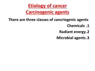
Examples of major chemicals: 1. Direct acting chemicals. - Alkylating agents like (anticancer drugs e.g. cyclophosphomides) - Acylating agents like (imidazole) 2. Indirect acting chemicals (procarcinogens) - Polycyclic aromatic hydrocarbons (like Benzanthracene ) - Aromatic amines, amides, azo dyes (like beta- naphthylamine) - Natural plants & Microbial products (Aflatoxin B1, Griseofulvin) - Others (Nitrosamines, Vinyl chloride, nickel, chromium, insecticides, fugosides, Asbestos)
Tumor Antigens: These are Antigens resent on the tumors that elicit the Immune response. These antigens are broadly divided into 1. Tumor specific Antigens: These only present on tumor cells & not on the normal cells. 2. Tumor associated Antigens: These antigens are present only on the tumor cells as well as on the normal cells. Examples of Tumor specific Antigens: 1. Viral antigens: These Antigens are derived from oncogenic virus like HBV, HPV. 2. Oncofetal Antigens: Like carcinoembryonic Antigens (CEA) & Alfa- Fetoprotein (AFP). These are present during embryogenesis but not in the normal adult tissue. Derepression of gene encodes these Antigens; this will result in Re-expression of these Antigens as in carcinoma of colon & liver.
3. Differentiated specific Antigens: Like Prostatic Specific Antigen (PSA), EXPRESS IN BOTH BENIGN & MALIGNANT PROSTATIC TISSUE (BUT IN DIFFERENT LEVEL). Effects of tumors on the host: Cancers are far more threatening to the host than benign tumors. Both benign & malignant affect the host. By the followings: 1. Location of tumors (benign & malignant) & their effects on adjacent tissue, even small size, benign tumor can cause problem to the host like pituitary adenoma less than 1cm can cause compression of adjacent tissue. 2. Effects on functional activity of the host: Both benign & well differentiated malignant tumors arising in endocrine
glands, e.g. adenoma & Carcinoma of adrenal gland cause increase level of steroid hormone that has effects on the host. 3. Producing bleeding & secondary infection, when the lesion is ulcerated through adjacent tissues (one of important cause of death in malignant tumors) 4. Many malignant tumor produce Cachexia & Paraneoplastic syndrome. Cancer Cachexia: It is referring to progressive loss of body fat, lean body mass, accompanied by profound weakness, anemia, & anorexia. There is correlation between the size & extent of spread of cancer & severity of cachexia, e.g. small size malignant tumor not produce Cachexia.
Pathogenesis of cachexia: It is of multifactorial pathognesis 1. Anorexia: It is common problem in patient with cancer; even in those don’t have cancer of GIT. So the cause of Anorexia is due to central cause like inhibition of taste & appetite center. 2. Increase Basal Metabolic Rate (BMR): In patient with cancer there is increase BMR & Calorie expenditure, the exact mechanism of this change is not fully understood, it is thought that, there is inhibition of appetite center by TNF-1 & IL-1 from activated macrophages. Also these factors cause inhibition of lipase enzyme, which result loss of release of free fatty acids fro lipoproteins. 3. Protein mobilizing factor has been detected in the serum of patient with cancer (skeletal muscle weakness). 4. Lipolytic Factor is thought to be the cause of Cachexia.
Paraneoplastic syndromes: Symptom complex other than Cachexia that occur in patient with cancer & that cannot be readily explained by local or distant spread of the tumor or by elaboration of hormones indigenous to tissue of origin of tumor. They appear in 10 – 15% of patient with cancer & it is important to recognize them for many reasons, include: 1. They may represent early manifestation of occult cancer. 2. In the affected patient, may represent significant problems & may be lethal. 3, they may mimic metastatic cancer.
Grading and Staging of Cancer: Methods to quantify the probable clinical aggressiveness of a given neoplasm and to express its apparent extent and spread in the individual patient are necessary for comparisons of end results of various forms of treatment. The grading of a cancer attempts to establish some estimate of its aggressiveness or level of malignancy based on the cytologic differentiation of tumor cells and the number of mitoses within the tumor. The cancer may be classified as grade I, II, III, or IV, in order of increasing anaplasia. Criteria for the individual grades vary with each form of neoplasia and so are not detailed here.
Staging of cancers is based on the size of the primary lesion, its extent of spread to regional lymph nodes, and the presence or absence of metastases. This assessment is usually based on clinical and radiographic examination (computed tomography and magnetic resonance imaging) and in some cases surgical exploration. Two methods of staging are currently in use: the TNM system (T, primary tumor; N, regional lymph node involvement; M, metastases) and the AJC (American Joint Committee) system. In the TNM system, T1, T2, T3, and T4 describe the increasing size of the primary lesion; N0, N1, N2, and N3 indicate progressively advancing node involvement; and M0 and M1 reflect the absence or presence of distant metastases. In the AJC method, the cancers are divided into stages 0 to IV, incorporating the size of primary lesions and the presence of nodal spread and of distant metastases.