Download
1 / 15
190 likes | 437 Views
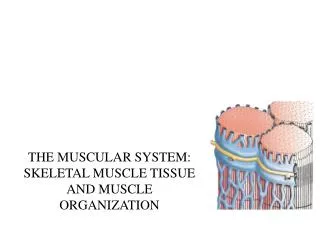
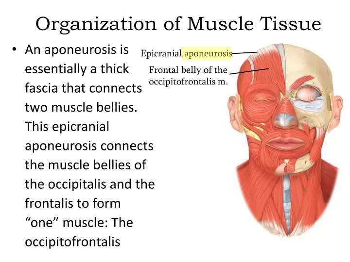
Organization of Muscle Tissue. Epicranial aponeurosis. An aponeurosis is essentially a thick fascia that connects two muscle bellies. This epicranial aponeurosis connects the muscle bellies of the occipitalis and the frontalis to form “one” muscle: The occipitofrontalis.

E N D
Organization of Muscle Tissue Epicranial aponeurosis • An aponeurosis is essentially a thick fascia that connects two muscle bellies. This epicranial aponeurosis connects the muscle bellies of the occipitalis and the frontalis to form “one” muscle: The occipitofrontalis Frontal belly of the occipitofrontalis m.
Organization of Muscle Tissue Veins, arteries, and nerves are located in the deep fascia between muscles of the thigh.
The Skeletal Muscle Fiber • Beneath the connective tissue endomysium is found the plasma membrane (called the sarcolemma) of an individual skeletal muscle fiber • The cytoplasm (sarcoplasm) of skeletal muscle fibers is chocked full of • contractile proteins • arranged in myofibrils
The Skeletal Muscle Fiber • You should learn the names of the internal structures of the muscle fiber • Sarcolemma • Sarcoplasm • Myofibril • T-tubules • Triad (with • terminal cisterns • Sarcoplasmic reticulum • Sarcomere
The Skeletal Muscle Fiber • Increasing the level of magnification, the myofibrils are seen to be composed • of filaments • Thick filaments • Thing filaments
The Skeletal Muscle Fiber • The basic functional unit of skeletal muscle fibers is the sarcomere: An arrangement of thick and thin filaments sandwiched between two Z discs A scanning electron micrograph of a sarcomere
The Skeletal Muscle Fiber • Muscle contraction occurs in the sarcomeres The “Z line” is really a Z disc when considered in 3 dimensions. A sarcomere extends from Z disc to Z disc.
Muscle Proteins • Myofibrils are built from three groups of proteins • Contractile proteins generate force during contraction • Regulatory proteins help switch the contraction process on and off • Structural proteins keep the thick and thin filaments in proper alignment and link the myofibrils to the sarcolemma and extracellular matrix
Muscle Proteins • The thin filaments are comprised mostly of the structural protein actin, and the thick filaments are comprised mostly of the structural protein myosin • However, in both types of filaments, there are also other structural and regulatory proteins
Muscle Proteins • In the thin filaments actin proteins are strung together like a bead of pearls • In the thick filaments myosin proteins look like golf clubs bound together
Muscle Proteins • In this first graphic, the myosin binding sites on the actin proteins are readily visible. • The regulatory proteins troponin and tropomyosin have been added to the bottom graphic: The myosin binding sites have been • covered
Muscle Proteins • In this graphic the troponin-tropomyosin complex has slid down into the “gutters” of the actin molecule unblocking the myosin binding site • The troponin-tropomyosin complex can slide back and forth depending on the presence of Ca2+ Myosin binding site exposed
Muscle Proteins • Ca2+ binds to troponin which changes the shape of the troponin-tropomyosin complex and uncovers the myosin binding sites on actin
Muscle Proteins • Besides contractile and regulatory proteins, muscle contains about a dozen structural proteins which contribute to the alignment, stability, elasticity, and extensibility of myofibrils • Titan is the third most plentiful protein in muscle, after actin and myosin - it extends from the Z disc and accounts for much of the elasticity of myofibrils • Dystrophin is discussed later as it relates to the disease of muscular dystrophy
The Sliding-Filament Mechanism • With exposure of the myosin binding sites on actin (the thin filaments)—in the presence of Ca2+ and ATP—the thick and thin filaments “slide” on one another and the sarcomere is shortened