Download
1 / 35
360 likes | 792 Views
Anatomy, diagnosis and classification of sports injuries in the shoulder. Mr. Nnamdi Obi Specialist registrar United Kingdom. Objectives. Review anatomy of the shoulder Review history and examination Acute traumatic shoulder instability. Introduction. Instability Glenohumeral dislocation

E N D
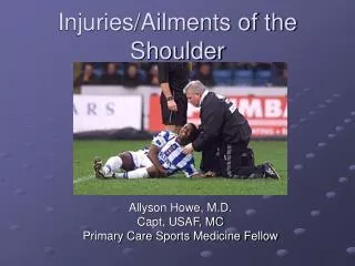
Anatomy, diagnosis and classification of sports injuries in the shoulder Mr. Nnamdi Obi Specialist registrar United Kingdom
Objectives • Review anatomy of the shoulder • Review history and examination • Acute traumatic shoulder instability
Introduction • Instability • Glenohumeral dislocation • SLAP tears • ACJ dislocation
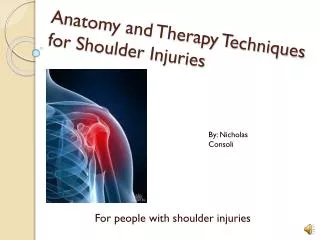
Anatomy • Synovial ball and socket joint • Articular surface covered with hyaline cartilage • Glenoid cavity deepened by labrum • Articulations
Rotator cuff • Supraspinatus • Infraspinatus • Teres Minor • Subscapularis
Ligaments • Glenohumeral • Superior Glenohumeral ligament • Middle Glenohumeral Ligament • Inferior Glenohumeral Ligament • Shoulder girdle • Coracoclavicular • ACJ proper • Acromioclavicular
Biomechanics Static restraints Dynamic restraints Rotator cuff muscles Biceps tendon Scapular stabilizers Neuromuscular factors • Glenoid labrum • Articular version + conformity • Glenohumeral ligaments • Negative intra-articular pressure
History(Acute traumatic instability) • Age • Mechanism • Traumatic • Atraumatic • Chronicity • Ease of dislocation • Expectations • Return to play
Examination • Acutely • Pain limits most • Pre and post axillary nerve function • Sensory • Motor • Delayed • Hyperlaxity – predisposing • Provocative tests • Labral pathology (SLAP tear)
Labrum (SLAP) • O’Brien’s
Labrum • Load & Shift
Special investigations • Bones • Glenoid • Head humerus • Soft tissues • Rotator cuff • Labrum Ultrasound – no labrum MRI X Ray CT scan CT arthrogram MRI arthrogram
Lateral radiographs • Posterior oblique scapular projection (“Neerlateral”, Neer 1970) • Produces considerable image overlap • Transthoracic (Vastamakiand Solonen1980) • Image overlap • Axial (Warrick 1965) • Requires shoulder abduction • Modified axial (Rockwood 1984) • Some shoulder abduction • Velpeau lateral (Wallace and Hellier 1983) • Patient needs to sit up • Apical oblique (Garth, Slappey and Ochs 1984)
This is posterior dislocation But outlines glenoid and humeral head J Bone Joint Surg [Br] l988;70-B:457-60.
Axial view Small Hills sachs Anterior glenoid Fine Almost normal AP Same patient Apical oblique Large Hills sachs Blunting anterior glenoid
Bone loss - Plain x-ray - CT - CT recon
Treatment How long ?
MRI study • IR Labrum off glenoid • ER tension rests on glenoid • Randomized 40 pts • Sling IR Vs ER • Recurrence • IR 6/20, 30% • ER 0/20 J Shoulder Elbow Surg 2003;12: 413-15
Premise • Younger = recurrent instability = immobilize longer • Older = stiffness = mobilize sooner • No benefit to immobilization in internal rotation > 1 week in pts under 30 yrs of age • Age of less than thirty years at time of injury predicts increased recurrence. • Best available evidence does show a clinical benefit to treatment in external rotation over conventional sling immobilization, but this advantage did not reach significance • BUT most ITOI J Bone Joint Surg Am. 2010;92:2924-33
Take Home • Reduce • Sling comfort • Discard in 1 week • Physiotherapy, strengthen dynamic stabilizers • Under 30 years, continue contact sport • Counsel recurrence rate • Consider surgery following first dislocation
SLAP Lesions • May be associated with dislocation but commonly due to pull on the arm, weightlifting, throwing, tackling • Symptoms – clicking, pain with overhead activities • Clinically – pain with eccentric biceps loading (e.g. going down on bench press)
Acromioclavicular joint (ACJ) injuries • Usually injured by a direct fall onto the point of the shoulder • Scapular forced downwards • Clinically, lateral end of clavicle prominent
Treatment • Non Operative • Grade 1-3 • Operative • Grade 4-6
Conclusions • Acute instability common in athletes • Glenohumeral • ACJ • High level of function • Early return to play • Axillary or modified axillary view • Apical oblique
References • Websites: • https://www.shoulderdoc.co.uk • https://www.orthobullets.com
The End Email: njco@hotmail.com