Download
1 / 2
20 likes | 150 Views
Biopsy port with cap. Video connection. Insertion tube. Suction port. Working channel. Eyepiece. Light transmitting glass fiber bundle. Control knob. Viewing glass fiber bundle. Light source and camera. Joanna Cheung Pui Ying 2004193805 Christine Ho Ka Man 2004032647

E N D
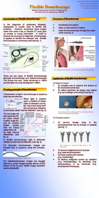
Biopsy port with cap Video connection Insertion tube Suction port Working channel Eyepiece Light transmitting glass fiber bundle Control knob Viewing glass fiber bundle Light source and camera Joanna Cheung Pui Ying 2004193805 Christine Ho Ka Man 2004032647 Veron Leung Ka Pik 2004125573 Eric Li Chun Hei 2004156871 Flexible BronchoscopeMedical Engineering, Faculty of Engineering, The University of Hong Kong, HKSAR Introduction to Flexible Bronchoscope In the diagnosis of pulmonary diseases, radiography is usually used to identify the problems. However, sometimes there maybe cases that chest x-ray or thoracic CT scan give us unclear or wrong information. In order to improve the accuracy of diagnosis, bronchoscopy is applied to identify the diseases and flexible bronchoscope is commonly used (see fig. 1). Procedure of Bronchoscopy • Anesthetize the patient. • Inject a small amount of saline. • Insert the bronchoscope through the nasal cavity (see fig. 5). Fig. 5 A flexible bronchoscope is used to view the airway of the patient[4] Fig. 1 Different parts of flexible bronchoscope [1] There are two types of flexible bronchoscope: fiberoptic bronchoscope and videobronchoscope. The former one was firstly introduced in 1960’s and the latter one was developed in 1990’s. • Application of flexible bronchoscope • For diagnostic purpose: • To visualize and to observe the lesions of the tracheobronchial tree • To collect specimen for biopsy and culture (e.g. by curettage or brushing) (see fig. 6) • For therapeutic purpose: • To remove foreign body in the tracheobronchial tree by forceps or baskets (see fig. 7) • 2. To remove malignant tumors by laser (e.g. laser photoresection) • 3. To frozen surface skin lesions (e.g. cryotherapy) • 4. To destroy malignant tumors by radiation (e.g. brachytherapy), by electricity (e.g. electrocautery) or by chemicals (e.g. photodynamic therapy)[3] Working principle of bronchoscope Transmission of light in bronchoscope is based on total internal reflection. When light is incident upon a medium of lesser index of refraction (ni>nt), theray is bent away from the normal such reflection 6a 6b Fig. 2 Internal reflection[2] is commonly called "internal reflection" (see fig. 2). Fig. 6a Brush used for collecting cells from tracheobronchial tree Fig. 6b Collecting cells by brushing[5] The exit angle will then approach 90° for some critical incident angle θc, and for incident angles greater than the critical angle there will be total internal reflection[2] (see fig. 3). Fig. 3 Total internal reflection[2] In flexible bronchoscope, light is delivered distally by total internal reflection. (see fig. 4) For fiberoptic bronchoscope, images are brought back to eyepiece using this principle, too. Fig. 7 Forceps for removing foreign body in the bronchus[6] Fig. 4 Transmission of light within a optical fibre[2] For videobronchoscope, images are brought back by a small charge-coupled device (CCD) sensor in the tip part. Better image resolution can be obtained[3].
1. Internal recording device • Allow the bronchoscopist to check the previous findings and operations. • Enhance patient management.2. More portable system • Install more portable components, say a smaller LCD monitor and a smaller image processing machine. (see fig. 10) • Autofluorescence bronchoscopy • Make use of the characteristics of autofluorescence on tumor to observe carcinoma or dysplasia using special light source and equipment[12] (see fig. 11). 11a 11b Fig. 10 A smaller LCD monitor is more portable and convenient.[11] Fig. 11a White light image. No abnormal area visible.Fig. 11bAutofluorescence image. Suspicious lung tissue with significantly reduced fluorescence.[13] Acknowledgements We thank Dr. Ian Gibson and Dr. Nelson Yung for giving the engineering information and Ms. Miranda Legg for the audio assistance. Special thanks to Dr. David Chong for showing us the demonstration of using flexible bronchoscope. [1] Emidicine consumer health website. Available at: http://www.emedicinehealth.com/. Accessed February 28, 2005. [2] HyperPhysics Concepts website. Available at: http://hyperphysics.phy-astr.gsu.edu/. Accessed March 1, 2005. [3] Bolliger C.T., Mathur P.N., ed. Interventional Bronchoscopy. Basel: Karger; 2000. [4] Your Medical Source website. Available at: http://yourmedicalsource.com/. Accessed February 28, 2005. [5] Tobacco Facts website. Available at: http://www.tobacco-facts.info/. Accessed March 1, 2005. [6] Untitled. Available at: http://www.hyunjoo.co.kr/. Accessed February 28, 2005. [7] Beamis J. F. Jr., Mathur P.N.. Interventional Pulmonology. The United States of America, the McGraw-Hill Companies, Inc.; 1999. [8] Karl Storz Veterinary Endoscopy America Inc. website. Available at: http://www.karlstorz.com/. Accessed March 1, 2005. [9] Cladonia Resources website. Available at: http://www.cladonia.co.uk/. Accessed March 2, 2005. [10] Beth Israel Deaconess Medical Center. Available at: http://bidmc.harvard.edu/. Accessed February 28, 2005. [11] LUXMA Manufacturer of Mobile Accessories website. Available at: http://www.luxmt.com/. Accessed March 1, 2005. [12] J.A. Nakhosteen, B. Khanavkar. Autofluorescence Bronchoscopy: The Laser Imaging Fluorescence Endoscope. In: Bolliger C.T., Mathur P.N., ed. Interventional Bronchoscopy. Basel: Karger; 2000:236-242. [13] American College of Chest Physicians website. Available at: http://www.chestnet.org/. Accessed February 28, 2005. References Analysis of flexible bronchoscope Fig.8 The turning degree of a bronchoscope[8] Fig.9 The large set of monitor[10] Overall, flexible bronchoscope is useful despite the disadvantages it has. It helps the doctors in visualizing the tracheobronchial tree and this allows a better diagnosis for pulmonary diseases. In addition, operations can be carried out through flexible bronchoscopy. Further development of flexible bronchoscope