Download
1 / 69
690 likes | 841 Views
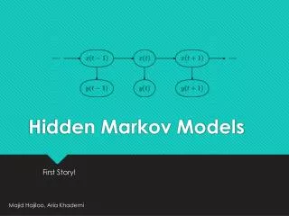
Hidden Markov Models. Probabilistic model of a Multiple sequence alignment. No indel penalties are needed Experimentally derived information can be incorporated Parameters are adjusted to represent observed variation. Requires at least 20 sequences. The Evolution of a Sequence.

E N D
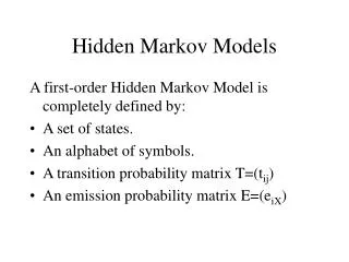
Hidden Markov Models • Probabilistic model of a Multiple sequence alignment. • No indel penalties are needed • Experimentally derived information can be incorporated • Parameters are adjusted to represent observed variation. • Requires at least 20 sequences
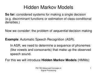
The Evolution of a Sequence • Over long periods of time a sequence will acquire random mutations. • These mutations may result in a new amino acid at a given position, the deletion of an amino acid, or the introduction of a new one. • Over VERY long periods of time two sequences may diverge so much that their relationship can not see seen through the direct comparison of their sequences.
Hidden Markov Models • Pair-wise methods rely on direct comparisons between two sequences. • In order to over come the differences in the sequences, a third sequence is introduced, which serves as an intermediate. • A high hit between the first and third sequences as well as a high hit between the second and third sequence, implies a relationship between the first and second sequences.Transitive relationship
Introducing the HMM • The intermediate sequence is kind of like a missing link. • The intermediate sequence does not have to be a real sequence. • The intermediate sequence becomes the HMM.
Introducing the HMM • The HMM is a mix of all the sequences that went into its making. • The score of a sequence against the HMM shows how well the HMM serves as an intermediate of the sequence. • How likely it is to be related to all the other sequences, which the HMM represents.
B M1 M2 M3 M4 E Match State with no Indels MSGL MTNL Arrow indicates transition probability. In this case 1 for each step
B M1 M2 M3 M4 E Match State with no Indels MSGL MTNL S=0.5 T=0.5 M=1 Also have probability of Residue at each positon
B M1 M2 M3 M4 E Typically want to incorporate small probability for all other amino acids. MSGL MTNL S=0.5 T=0.5 M=1
B M1 M2 M3 M4 E Permit insertion states MS.GL MT.NL MSANI I0 I1 I2 I3 I4 Transition probabilities may not be 1
B M1 M2 M3 M4 E Permit insertion states MS..GL MT..NL MSA.NI MTARNL I0 I1 I2 I3 I4
DELETE PERMITS INCORPORATION OF LAST TWO SITES OF SEQ1 MS..GL-- MT..NLAG MSA.NIAG MTARNLAG AA GN IL ST A M D1 D2 D3 D4 D5 D6 D7 I7 I0 I1 I2 I3 I4 I5 I6 M4 E M2 M3 M5 M6 B M1 M7 G
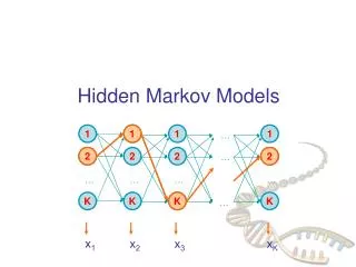
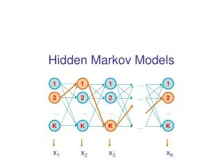
D1 D2 D3 D4 D5 D6 I0 I1 I2 I3 I4 I5 I6 B M1 M2 M3 M4 M5 M6 E • The bottom line of states are the main states (M) • These model the columns of the alignment • The second row of diamond shaped states are called the insert states (I) • These are used to model the highly variable regions in the alignment. • The top row or circles are delete states (D) • These are silent or null states because they do not match any residues, they simply allow the skipping over of main states.
Dirichlet Mixtures • Additional information to expand potential amino acids in individual sites. • Observed frequency of amino acids seen in certain chemical environments • aromatic • acidic • basic • neutral • polar
STRUCTURES a helix b sheet coils turns Structures are used to build domains.-Legos of evolution
Ramachandran plot for Glycine Areas not permitted for other amino acids Psi Angles Phi angles
Introduction to Protein Structure, Branden and Tooze Garland Publishing Co.1991 p.13
From: http://bioweb.ncsa.uiuc.edu/~bioph254/Class-slides/Lect12/figure13.html
Longitudinal and Transverse image of alpha helix From: http://bioweb.ncsa.uiuc.edu/~bioph254/Class-slides/Lect12/figure14.html
Turn connecting two helices Introduction to Protein Structure, Branden and Tooze Garland Publishing Co.1991 p. 17
Proline • Because of its structure, proline is typically excluded from a helices except in the first three positions at the amino end.
b Structure b strand - single run of amino acids in b conformation b sheet- multiple b strands which are hydrogen bonded to yield a sheet like structure. b bulge - disruption of normal hydrogen bonding in a b sheet by amino acid(s) that will not fit into the sheet -for example: proline
b sheets- Parallel Introduction to Protein Structure, Branden and Tooze Garland Publishing Co.1991 p.17.
b sheet - longitudinal and transverse view. Side chains stick “out” http://bioweb.ncsa.uiuc.edu/~bioph254/Class-slides/Lect12/figure22.html
Six classes of structure • Class a- bundled a helices connected by loops. • Class b- sandwich or barrel comprised entirely of b sheets typically anti-parallel. • Class a / b mainly parallel b sheets with intervening a helices. • Class a + b - segregated a helices and anti-parallel b sheets • Multi-domain • Membrane proteins
Endonuclease Class a + b
Rhodopsin 7TM proten
Common Hairpin Loop between two b Strands Introduction to Protein Structure, Branden and Tooze Garland Publishing Co.1991 p. 17
Turn - short, regular loop. • Difference in frequency of amino acids at positions 1-4 of the turn. • Coils (not coiled coil) • Random turns or irregular structure.
Disulfide bridges • Crosslink of two cysteine residues. • Distance between sulfur = 3 Angstroms.
Coiled coil -two a helices bundled side by side From: http://catt.poly.edu/~jps/coilcoil.html
a,d are internal, remaining amino acids are solvent exposed From: http://catt.poly.edu/~jps/coilcoil.html
Coiled Coil • Two or more adjacent a helices
Potential Residues involved in Coiled Coil MMFPQSRHSGSSHLPQQLKFTTSDSCDRIKDEFQLLQAQYHSL KLECDKLASEKSEMQRHYVMYYEMSYGLNIEMHKQAEIVKR LNGICAQVLPYLSQEHQQQVLGAIERAKQVTAPELNSIIRQQL QAHQLSQLQALALPLTPLPVGLQPPSLPAVSAGTGLLSLSALG SQTHLSKEDKNGHDGDTHQEDDGEKSD
Domains • Single domain proteins - • Epidermal growth factor • Serine Proteases - Trypsin • Multi domain proteins -Factor IX -one Ca2+ binding, two EGF/ one protease domain. • Permit building of novel functions by swapping of domains
Factor IX Domain Structure Ca EGF EGF CT Ca - Calcium binding domain EGF - Epidermal growth factor domain CT - Chymotrypsin domain
Chou - Fasman Prediction of Secondary Structure • Based upon analysis of known structures (1974). • Frequency of occurrence of each amino acid in: • a helix • b strand • turn
Chou - Fasman Prediction • List is then analyzed for stretches of amino acids that have a common tendency to form a given secondary structure. • Extend until a region of high probability for either a turn or region with a low probability of both a or b is encountered. • Window is typically <10