Download
1 / 54
610 likes | 1.11k Views
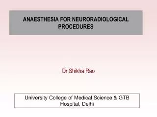
ANAESTHESIA FOR NEURORADIOLOGICAL PROCEDURES. Dr Shikha Rao. University College of Medical Science & GTB Hospital, Delhi. Common Neuroradiological Procedures. Radiology Computed tomography Magnetic resonance imaging Interventional neuroradiology (vascular & non vascular)

E N D
ANAESTHESIA FOR NEURORADIOLOGICAL PROCEDURES Dr Shikha Rao University College of Medical Science & GTB Hospital, Delhi
Common Neuroradiological Procedures Radiology • Computed tomography • Magnetic resonance imaging • Interventional neuroradiology (vascular & non vascular) • Functional brain imaging • Positron emission tomography
ASA Guidelines for nonoperating room anaesthetizing location Oxygen • Reliable source , type E full cylinder Suction • Adequate and reliable Scavenging system if inhalational agents are used Anaesthetic equipment • Self inflating bag to deliever IPPV • Adequate drug supply • Anaesthesia machine with equivalent standards to that in OT and maintained to same standards • Adequate monitoring equipments to allow adherence to ASA standards for basic monitoring
Electrical outlets • Sufficient for anaesthesia machine and monitors Adequate illumination • Battery operated backups Sufficient space for • Personnel and equipments • Easy and expeditious access to patient , anaesthesia machine, and monitoring Resuscitation equipment immediately available • Defibrillator • Emergency drugs • Cardiopulmonary resuscitation equipments
Adequately trained staff to support anaesthesia team All building and safety codes and facility standards should be observed Post anaesthesia care facilities • Adequately trained staff to provide post anaesthesia care • Appropriate equipment to allow safe transport to main postanaesthesia care unit
Patients requiring sedation or general anaesthesia for neuroradiology • Anxiety and panic disorders • Claustrophobia • Developmental delay and learning difficulties • Cerebral palsy • Seizure disorders • Movement disorders • Severe pain • Acute trauma with unstable cardiovascular, respiratory, or neurologic function • Significant comorbidity • Pediatric age group
Monitoring • Universal requirement • Basic monitoring standards Standard I requires a qualified anesthesia personnel to be present in the room throughout the conduct of anesthesia. Standard II -continous evaluation of the patient's oxygenation, ventilation, circulation, and temperature. Continuous Monitoring • ECG – heart rate & adequacy of circulation • NIBP – every 5 mins , adequacy of circulation
Pulse oximetry – oxygenation , measures pulse rate &estimate oxygen saturation of hemoglobin in a noninvasive & continuous fashion • Ventilation – assessed by observation & auscultation • End tidal CO2 - in intubated patients • side stream sampling • alteration in ventilation, cardiac output • Temperature • Radiology suites are often maintained at low temperatures for the computer system. • fluid warmer can be used to maintain patient’s body temperature
Anaesthetic techniques • Techniques vary from no anaesthesia to minimal or deep sedation (MAC) to general anaesthesia • It depends on • patient’s medical condition • desired level of anaesthesia • procedure to be performed • duration of procedure
Monitored anaesthesia care • Administration of drugs with anxiolytic, hypnotic, analgesic & amnestic properties either alone or in combination with local or regional anaesthesia Preoperative assessment • Detailed history & examination of patient (similar to that done before GA) + • Evaluation of ability of patient to remain motionless & if necessary actively cooperate throughout out procedure + • Fasting status
Following drugs are used in MAC Propofol • Sedative, hypnotic • Short Context sensitive half life, short effect site equilibration time • Rapid & clear headed recovery • Decreases incidence of nausea & vomiting • less incidence post procedure sedation , drowsiness • Has anti emetic properties (Subhynotic dose of 10 mg is said to be effective )
Benzodiazepines • Anxiolytic, amnestic & hypnotic properties • Midazolam • Commonly used for moderate to deep sedation • Short elimination half life( 1 to 4hrs) • Dose – 0.02 to 0.1 mg/kg • Diazepam • Longer elimination half time ( > 20 hrs) • active metabolites (desmethyl diazepam, oxazepam) • Flumazenil • should be used cautiously in benzodiazepine toxicity
Opioids • Provide analgesia component in balanced anaesthesia technique • Disadv - do not provide amnesia Adverse effects • Respiratory depression • muscle rigidity nausea / vomiting Sedation / anaesthetic drug interaction • Depends on combination, dose range over which administered • Opioids + benzodiazepines – synergism in hypnotic / analgesic / amnesic properties
General anaesthesia technique Required when the procedure involves • Many dermal punctures • Longer duration • Interventional neuroradiology General anaesthesia achieved using • Induction of GA done in side room or operating room & patient is transported to required facility • Anaesthesia machine should be kept near the patients feet, & opposite the neuroradiologist
Endotracheal intubation & IPPV or • Laryngeal mask airway with spontaneous breathing / IPPV • Induction of general anaesthesia – Propofol / Thiopentone • Maintenance of anaesthesia – volatile anaesthetics / TIVA • At the end of procedure patient is transported to recovery area where further management is provided by trained anaesthesia personnel
Radiology suite showing necessity for anaesthesia equipment and anesthesiologist to be remote from the patient's head
Computed Tomography (CT) • CT is a medical imaging method in which a 3-d image of inside of an object is generated from a large series of 2-d images taken around a single axis of rotation • Used for diagnostic or therapeutic purposes • Hypodense (dark) structures – infarction • Hyperdense (bright) - calcification &hemorrhage
Problems faced by anaesthesiologist • Inaccessibility of patient • Interference in monitoring • In an intubated patient, care should be taken that sides of scanning tunnel do not dislodge the circuit • Adverse effects of contrast media • Exposure to ionizing radiation
Patient monitoring • Basic monitoring standards • Monitors should be easily viewed Anaesthetic considerations Elective Emergency • Preanaesthetic check up on day of procedure • Head injury patients with ongoing blood loss or raised ICP • Considered full stomach patients • Either scanned awake or intubation following rapid sequence induction
Contrast media • Contrast media often used in CT scan • 2 types • Iodinated (I-53) – hyperosmolar & toxic, eg:Na iothalamate • Non iodinated – low osmolality & fewer side effects ,eg:iopamidol • Adverse effects • direct toxicity • idiosyncratic reactions • anaphylactic/ anaphylactoid reactions
Treatment –Usually symptomatic • O2 • Epinephrine • Bronchodilators • Corticosteroids role in biphasic anaphylaxis • Patients with past h/o of reaction to contrast media • Prednisolone 50 mg i.v. 12 & 2 hrs prior to the procedure • Diphenhydramine 50mg immediately before the procedure
Contrast induced nephropathy • Acquired ARF • Incidence high with hyperosmolar agents • For Prevention maintain proper hydration before, during & after the procedure • Usually self limited & resolve in 2 weeks • Acetyl Cysteine / Ascorbic acid MOA ? –antioxidant enhance vasodilatory effects of NO
Use suggested for Prophylaxis in high risk patients , suffering from diabetics , hypertension , CHF etc . Acetycysteine - 600 – 1200 mg orally twice daily for 2 doses before procedure & 2 doses after procedure Ascorbic acid -3gm p.o 2 h before procedure & 2gm daily twice daily the day after procedure (Pannu N,Wiebe N , Tonelli M.prophlaxis strategies for contrast induced nephropathy. JAMA,June 21, 2006- Vol295,No.23)
Radiation exposure • More with CT scan than any other radiological procedure • Radiation toxicity • Somatic effects • Genetic injury • Dosimeters monitor exposure • Maximum permissible exposure • 50 mSv /year • Cumulative lifetime dose of 10mSv x age • 0.5 mSv for pregnant women
Ionizing Radiation follow Inverse square law – radiation exposure decreases proportional to square of distance from the source • Radiation exposure – limited by • lead aprons • thyroid shields • using movable leaded glass screens – anaesthesiologist can stand across the screen & monitor the patient
Magnetic resonance imaging • Can differentiate areas of dissimilar anatomy, physiology & pathology • Noninvasive , no ionizing radiation used • Provides excellent soft tissue contrast • Can obtain image in any plane • Differentiates b/w white & grey matter, permits resolution of CSF flow
Disadvantages • Calcium does not emit any signal – prevents detection of pathologic calcification in soft tissues • Time consuming • Patient Movement can produce artifact • Noise > 90db
Zone Definition • Zone 1 – includes all areas freely accesible to general public i.e area outside MRI enviornment • Zone II – area b/w publicly accessible zone 1 & strictly controlled zone III ,patients are greeted in zone II but movement is supervised by MR personnel • Zone III- access to this zone strictly restricted & controlled be MR pesonnel . No ferromagnetic object should be brought in zone • Zone IV- MR scanner magnet room NOTE - At the 30-50 gauss line( Zone II), ferromagnetic equipment may be used safely.. (Jorgensen, et.al. 1994)
Categories of patient requiring anaesthesia or sedation in MRI
Concerns in anaesthetic management • Patient access & visibility • Absolute need to exclude ferromagnetic component • Interference / malfunction of monitoring equipments produced by changing magnetic fields • Potential degradation of imaging caused by stray radiofrequency currents from monitoring equipments • Limited access to MRI suite • Potential for heat generation within monitoring wires as a result of electro magnetic conduction
Recommendations to prevent thermal injury • Avoid loop formation of monitoring wires, keeping them straight • Avoid conductors touch the patient at more than one location • Inspection of monitor wires before every use
To avoid above listed problems –anaesthesiologist must be involved in planning & construction of MRI suites • Common approach • In case a patient needs anaesthesia, it is induced outside MRI room & patient transported on non ferromagnetic tables • For emergence or in case of emergency, patient brought out on same table
Monitoring equipment requirements for MRI • ECG – liquid crystal screens, high impedance graphite electrodes & leads avoid loops of wire • Blood pressure- oscillometer with nonferrous guage • Respiratory gas – side stream sampling with long sampling line • Temperature – skin temperature sensing strips (burns reported with probes) • Pulse oximeter – non-ferromagnetic model
MRI compatible anaesthetic equipment • Laryngoscope – plastic scopes with paper or aluminium covered lithium cells • Stylet – copper model available • Endotracheal tube – spring within valve cuff may distort image; nonmagnetic version is available, avoid metal reinforced tubes & metal connectors • Laryngeal mask airway - spring within valve cuff may distort image ;nonmagnetic version is available • Ventilator – available
Anaesthetic machine – nonmagnetic machine, aluminium cylinders required , breathing circuits (Aestiva5 MRI workstation from Datex Ohmeda) • Infusion pumps – used at 30 gauss line , but extensions recommended to minimize field effect on motor function • Self inflating bags – valveless with no magnetic parts • Suction – wall mounted with a 10 m tubing • Defibrillators – resuscitation usually carried out outside magnetic field
Implanted devices or objects representing a contraindication to MRI • Cardiac pacemaker • Metal eye splinter or shrapnel • Vascular clips or intrauterine contraceptive devices • Interventional radiology device • Orthopedic device (prosthetic joint , wire plate)
Anaesthesia for interventional neuroradiological procedures Procedures can be diagnostic & therapeutic, like • Embolization of cerebral & dural AVM • Coil embolization of cerebral aneurysms • Thrombolysis of thromboembolic stroke • Awake craniotomy
Real time imaging • Digital subtraction angiography • High resolution flouroscopy • Contrast media used • Radiation exposure more with DSA than flouroscopy Vascular access • Femoral / carotid / brachial artery
Anaesthetic Considerations • Maintanence of patient’s immobility • Physiologic stability –Manipulation of regional & systemic blood flow • Evaluation of coagulation profile • Treating complications that can occur during the procedure • Rapid transition b/w sedation & awake responsive state
Anaesthetic management • Preprocedure anaesthetic evaluation –careful neurological examination ( preexisting deficits & Glasgow Coma Score) • Airway examination • as airway evaluation during the procedure not possible because of head position, better to secure airway early in patients at risk of airway compromise • Standard anaesthesia monitoring established • Two intravenous access established • Invasive arterial blood pressure monitoring - radial artery • Deliberate hypotension • During embolization of AVM • Drugs used are esmolol, SNP, labetolol
Deliberate hypertension induced cerebral ischemia • To increase collateral flow • Drugs – phenylephrine, vasopressin • Close ECG monitoring done to detect signs of myocardial ischemia • Urinary catheter – use of large amounts of radiologic contrast media & osmotic diuretic agents • Sedation – combination of BZD & opioids • GA – accomplished by volatile or TIVA (considering desirability of emergence for neurologic evaluation) • Padding of pressure points • Antiemetic should be given
Embolization of cerebral aneurysms Carotid occlusion test- to test for adequacy of cerebrovascular collateral circulation Wada test -behavioural testing done after injection of sodium methohexital into ICA in an awake patient , done to determine dominant site for vital cognitive function Superselective anaesthesia functional examination( SAFE)
Interventional MRI • In awake craniotomies for accurate tumour resection • Horse shoe shaped scanners used • Follow sleep – awake – sleep cycle • Titanium instruments used • Patient access limited
Complications of interventional neuroradiological procedures CNS – Haemorrhagic – • aneurysm perforation • intracranial vessel injury Occlusive – • thromboembolic phenomenon • displacement of coil • vasospasm OTHERS- Contrast reaction Contrast nephropathy Haemorrhage at puncture site , groin hematoma
Management of complications • Initial resuscitation • Communicate with radiologist • Call for assistance • Secure the airway & ventilate with 100% O2 • Determine whether problem is haemorrhagic or occlusive • If haemorrhagic- Immediate heparin reversal, Delibrate hypotension • If occlusive- Delibrate hypertension • Head up position • Mannitol to be given i/v • Consider dexamethasone , anticonvulsants – dilantin
Special considerations Pediatric Patients An anaesthesiology referral required in pediatric patients with the following conditions before any procedure • h/o apnoea • Age < 1 yr • Respiratory compromise • Pierre robin syndrome • Aperts / crouzon’s syndrome • Severe gastrointestinal reflux • Poor oral muscle development • Sedation / analgesic allergy • New onset illness • Cardiac disease • Mitochondrial / metabolic illness • Cerebral palsy (Children Hospital of Boston)