Monolocular Scalloped Radiolucency in Teeth 35 to 37: Clinical Insights
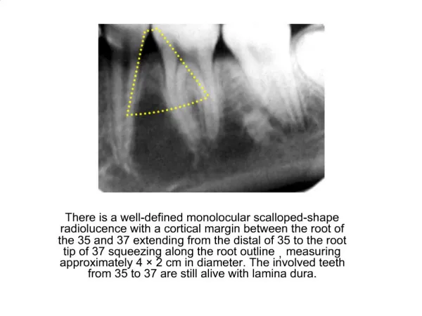
A well-defined monolocular scalloped radiolucency presents with a cortical margin between the roots of teeth 35 and 37, extending from the distal of tooth 35 to the root tip of tooth 37. This lesion measures approximately 4.2 cm in diameter and follows the contour of the root outline. Notably, the affected teeth remain vital, with intact lamina dura. This finding raises questions about the etiology and clinical management of the radiolucency while highlighting the importance of careful radiographic evaluation.

Monolocular Scalloped Radiolucency in Teeth 35 to 37: Clinical Insights
E N D
Presentation Transcript
1. There is a well-defined monolocular scalloped-shape radiolucence with a cortical margin between the root of the 35 and 37 extending from the distal of 35 to the root tip of 37 squeezing along the root outline,measuring approximately 4 � 2 cm in diameter. The involved teeth from 35 to 37 are still alive with lamina dura.