Download
1 / 51
1.33k likes | 2.68k Views
Histology of Female Reproductive System. Petek Korkusuz MD PhD. Aim. To learn the histology and the histophysiology of female genital system. Learning Goals. To learn the histology and histophysiology of the ovaries To learn the histology and histophysiology of the oviducts

E N D
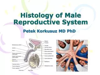
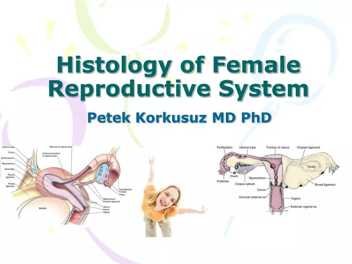
Histology of Female Reproductive System Petek Korkusuz MD PhD
Aim • To learn the histology and the histophysiology of female genital system
Learning Goals • To learn the histology and histophysiology of the ovaries • To learn the histology and histophysiology of the oviducts • To learn the histology and histophysiology of the uterus • To learn the histology and histophysiology of the vagina • To learn the histology and histophysiology of the external genitalia
Female genital system • Internal reproductive organs • paired ovaries • oviducts, • Uterus • vagina • External genitalia (vulva) • Mons pubis • Labia majora • Labia minora • Vestibule • clitoris
Ovaries • Located within the pelvis, are almond-shaped bodies 3 cm long, 1.5 to 2 cm wide, and 1 cm thick. • Surface epithelium covering the ovaries is called the germinal epithelium(simple squamous low cuboidal epith), is a modified peritoneum. Directly beneath this epithelium is the tunica albuginea, the connective tissue capsule whose collagen fibers are oriented parallel to the ovary surface. • Each ovary is subdivided into the highly cellular cortex and a medulla.
Ovaries • Ovarian cortex is composed of a connective tissue framework, the stroma, housing fibroblast-like stromal cells,ovarian follicles in various stages of development. • Medulla contains interstitial cells, large blood vessels, lymph vessels, and nervefibers embedded in a connective tissue stroma.
Ovaries • Before the onset of puberty, all of the follicles of the ovarian cortex are in theprimordial follicle stage. • The pulsatile release of GnRH from the hypothalamus results in a similar, pulsatile, release of gonadotropins [FSH], and [LH]) from the basophils of the anterior pituitary that culminates in the commencement of follicular development and the onset of the ovulatory cycle(follicular-ovulatory-luteal phase).
Ovarian follicles • The development of the primary follicles is independent of FSH; differentiation and proliferation of the follicular cells are triggered by local factors secreted by cells of the ovary. • Secondary and later folliclesare under the influence of FSH. • Follicular development usually culminates in the release of a single oocyte (ovulation).
Ovarian follicles • Are surrounded by stromal tissue • Consist of a primary oocyte and its associated follicular cells arranged in a single spherical layer or several concentric layers around the primary oocyte. • There are 4 stages of follicular development based on the growth of the follicle and the development of the oocyte: • Primordial follicles • Unilaminarand multilaminar primary follicles • Secondary (antral)follicles • Graafian(mature)follicles
Primordial follicles • The most primitive follicles, • Retained in a resting phase in fetal ovary • Are abundant before birth, after which they become fewer in number. • Is composed of a primary oocyte, arrested in the prophase stage of meiosis I • is surrounded by a single layer of flattened follicular cells.
Primary follicles • Primordial follicles develop intoprimary folliclesdistinguished as a result of changes in the primary oocyte, the follicular cells, and the surrounding stromal tissue. • The primary oocyte grows to100-150 μm in diameter with an enlarged nucleus (sometimes called the germinal vesicle). • Follicular cells become cuboidal in shape. As long as only a single layer of follicular cells encircles the oocyte, the follicle is called a unilaminar primary follicle.
Multilaminar primary follicles • Follicular cells proliferate and stratify, forming several layers of cells around the primary oocyte, the follicle is called a multilaminar primary follicle, and the follicular cells are more commonly referred to as granulosa cells. • During this stage, an amorphous substance (the zona pellucida) appears, separating the oocyte from the surrounding follicular cells. • Microvilli of the oocyte and filopodia of the follicular cells invade the zonula pellucida and form gap junctions through which they communicate throughout follicular development.
Multilaminar primary follicles • Stromal cells form • an inner theca interna, composed mostly of a richly vascularized cellular layer • an outer theca externa, composed mostly of fibrous connective tissue. • Theca interna cells produceandrostenedionewhich enters the granulosa cells, where it is converted by the enzyme aromatase into the estrogen estradiol. • Granulosa cells are separated from the theca interna by a thickened basal lamina.
Secondary follicles • Are similar to primary follicles except for the presence of accumulations of liquor folliculi(containing hyaluronate, growth factors, gonadotropins) among the granulosa cells. • Continued proliferation of the granulosa cells of the secondary follicle depends on FSH released by basophil cells of the anterior pituitary. • As more fluid is produced, individual droplets of liquor folliculi coalesce to form a single, fluid-filled chamber, the antrum.
Secondary follicles • Granulosa cells become rearranged so that the primary oocyte is now surrounded by a small group of granulosa cells that project out from the wall into the fluid-filled antrum. This structure is called the cumulus oophorus. • The loosely arranged low cuboidal granulosa cells immediately adjacent to the ZP move slightly away from the oocyte, but their filopodia remain within the ZP, maintaining contact with the primary oocyte. • This single layer of granulosa cells that immediately surrounds the primary oocyte is called the corona radiata.
Preantral follicle formed by an oocyte and several layers of granulosa cells. The oocyte is surrounded by the zona pellucida Antral follicle
Secondary follicles • Most of the follicles that reach this stage of development undergo atresia. • A few secondary follicles continue to develop into mature follicles.
Graafian follicles • Continued proliferation of the granulosa cells and continued formation of liquor folliculi result in the formation of a graafian (mature) follicle • GF diameter reaches 2.5 cm by the time of ovulation. • GF may be observed as a transparent bulge on the surface of the ovary, nearly as large as the ovary itself. • The follicular cells of the wall of the follicle compose the membrana granulosa.
Graafian follicles • Continued formation of liquor folliculi causes the cumulus oophorus composed of the primary oocyte, the corona radiata, and associated follicular cells to become detached from its base to float freely within the liquor folliculi. • By14th day of the menstrual cycle, estrogen produced mostly by the developing graafian follicle, but also by secondary follicles, causes elevation of blood estrogen to levels high enough to shutting off of FSH release and a surge in LH release.
Graafian follicles-ovulation • The high blood levels of LH causes the completion of the first phase of meiosis I, resulting in the formation of the secondary oocyte. • The secondary oocyte begins, and is arrested in, the metaphase stage of meiosis II and is released from the graafian follice, a process known as ovulation. • The remnants of the graafian follicle are converted into the corpus hemorrhagicum and then the corpus luteum. Corpus luteum. Granulosa lutein cells constituting the majority of the cells, derive from the granulosa layer. They are larger and stain more lightly than the theca lutein cells originating from the theca interna.
Corpus luteum • Formed from the collapsed follicle wall that contains the granulosa and theca cells • Granulosa lutein cells form a thick folded layer around the former follicular cavity (cav) • Wthin the folds are cells of theca interna (arrows) • High magn shows the wall of CL mainly composed of granulosa lutein cells with rounded nucleus. • The theca lutein cells also have a rounded nucleus, but they are smaller than granulosa lutein cells.
A small portion of a corpus luteum. Most cells present in the figure are granulosa lutein cells.
Photo micrograph corpus albicans: large amounts of hyaline material can be seen among degenerating cells of the former CL. Electron micrograph of theca lutein cells of CL
Ovaries Scanning electron micrograph of an ovary, showing an oocyte surrounded by follicular cells. The structure covering the oocyte is the zona pellucida, which appears as an irregular meshwork. x2950.
Hypothalamo-hypophiseal-ovarian axis • Pituitary hormones control most ovarian functions. • FSH stimulates follicular growth and synthesis of estrogen by the granulosa cells. • LH induces ovulation and transforms the granulosa layer and the theca interna into an actively secreting gland, the corpus luteum. • Estrogen and progesterone produced in the ovary act on the hypothalamus, stimulating or inhibiting the liberation of GnRH.
Uterine (Fallopian) tubes • Paired tubes extending bilaterally from the uterus toward the ovaries • Transport ovum from the ovary to uterus; provide necessary envitonment for fertilization and initial development of zygote to the morula stage • One end of the tube is adjacent to the ovary and opens into peritoneal cavity; the other end communicates with the uterine cavity • Each tube is approx 10-12 cm long and divide into 4 segments: • İnfindibulum with fimbria, • ampulla, • isthmus, • uterine (intramural) part
Uterine (Fallopian) tube wall is composed of three layers: • Serosa (or peritoneum): outermost layer composing of mesothelium and a thin layer of connective tissue • Muscularis: thoughout most of its length is made of an inner, realitevly thick circular and an outer thinner longitudinal layer. The boundary between these layers is often indistinct • Mucosa: inner lining consisting of thin longitudinal fols that project into the lumen of the uterine tube. Folds are most numerous and complete in ampulla, become smaller in isthmus. Epith is simple columnar composing of cilliated and noncilliated (peg) cells.
Uterine (Fallopian) tube wall • 2 different epith cell types represent different functional states of a single cell type. • The epith cells undergo cyclic hypertrophy during follicular phase and atrophy during the luteal phase inresponse to changes in hormonal levels particularly estrogen. • The uterine tube demonstrates active movements just before ovulation as the fimbriae become closely apposed to the ovary and localize over the region of the ovarian surface where rupture will occur.
Uterus • A single, thick, pear-shaped structure located in the midline of the pelvis, • Receives at its broad, closed end the terminals of the paired oviducts. • It is divided into three regions: • body, • fundus, • cervix. • The uterine wall of the body and the fundus is composed of • endometrium • myometrium • adventitia or serosa.
Endometrium • Mucosal lining of the uterus, • is composed of a simple columnar epithelium and a lamina propria. • Epithelium is composed of nonciliated secretory columnar cells and ciliated cells, • Lamina propria houses simple branched tubular glands that extend as far as the myometrium • Morphological/ physiological alterations in the endometrium during the phases of the menstrual cycle are controlled by hormones.
Endometrium • Consists of two layers, • the E. functionalis, a thick, superficial layer that is sloughed at menstruation • the E. basalis, a deep, narrow layer whose glands and connective tissue elements proliferate and regenerate the functionalis during each menstrual cycle. • E.functionalis is vascularized by numerous coiled helical arteries that supply the glands and connective tissue. • The straight arteries are much shorter and supply only the E. basalis.
Myometrium-serosa • Myometrium is composed of inner longitudinal, middle circular, and outer longitudinal layers of smooth muscle. • Much of the anterior portion of the uterus is covered by adventitia , • Fundus and posterior portion of the body are covered by a serosa.
Menstruel cycle-menstruation • Menstbegins on the day bleeding from the uterus • Occurs when fertilization does not take place. • Corpus luteum becomes nonfunctional about 14 days after ovulation, thus reducing the levels of progesterone and estrogen. • Although the entire functionalis layer of the endometrium is sloughed, it is not completely released from the wall immediately; rather, this process continues for 3 to 4 days.
Menstruel cycle- proliferative (follicular) phase • FP occurs at the same time as the development of the ovarian follicles. • It begins when the menstrual flow ceases, on about day 4, • It continues through day 14 by which time the functionalis layer of the endometrium has been fully restored to its previous status with a full complement of epithelium, glands, stroma, and coiled arteries.
Menstruel cycle- secretory(luteal) phase • SP commences after ovulation. • During this phase, the endometrium continues to thicken. • Secretory products first accumulate in the basal region of the cytoplasm of the cells constituting the endometrial glands, • Granules move apically, and are released into the lumen of the gland. • This glycogen-rich material will nourish the conceptus before the formation of the placenta.
Menstruel cycle- secretory(luteal) phase • SP completes the menstrual cycle as the 28th day approaches • presaging the menstrual phase of a new menstrual cycle.
Cervix • Endometrium of the cervix is different from the rest of uterus • Mucosa measures 2-3 mm thick; containd large branched glands, lacks spiral arteries • Mucosa undergoes little change in thickness during menstr cycle, is not sloughed during menstruation. • Cervical glands undergo functional chnges realted to the transport of the spermatozoa • Amount and properties of the mucus secreted by the gland cells vary during the mens cycle. • At midcycle the amount of mucus increase 10 fold. • Blockage of opennings of mucosal glands results in the formation of nabothian cysts.
The transformation zone is the site of transition between vaginal startified squamous epith and cervical simple columnar epithelium.
Vagina • Is a fibromuscular tube that joins internal reproductive organs to the external environment • Extends from the cervix to the vestibule (area between labia minora) • In a virgin the opening into the vagina may be surrounded by the hymen • Hymen consists of folds of mucous membrane extending into the vaginal lumen
Vagina Vaginal wall consists of Inner mucosal layer: • with numerous transverse folds or rugae and is lined by startified squamous nonkeratinized epithelium. • CT papillae from the underlying lamina propria project into the epithelium, lacking glands. LP has two regions: outer cellular loose CT and deeper dense CT that may be considered as submucosa • Keratohyalin granules may be present in the epithelial cells, but keratinization does not occur
Vaginal epithelium • Epith surface is lubricated mainly by mucus produced by cervical glands and additionaly by vestibular glands • Epithelium undergoes cyclic changes during menstruel cycle: • Epith cells synthesize and accumulate glycogen as they migrate toward the surface during the follicular phase with estrogen • Epith cells continously are desquamated • Near or during the menstruel phase the superficial layer may be shed
Vagina Intermediate muscular layer • An outer longitudinal and inneer circular smooth muscle layer • The outer sm layer is continuous with corresponding layer in the uterus • Striated muscle fibers of the bulbospongiosus muscle are present at the vaginal opening Outer adventitial layer • organized into inner dense connective tissue layer adjacent to the muscularis • and an outer loose connective tissue
External genitalia(vulva) • Mons pubis • Labia majora • Labia minora • Vestibule • Clitoris
2 folds of skin heavily endowed with adipose tissue and a thin layer of smooth muscle; extending from mons pubis Homologue to scrotum Smooth muscle corresponds to dartos muscle of the scrotum Outer surface covered by pubic hair;devoid of hair on the inner surface Numerous sweat and sebaceous glands open on both surfaces Is the rounded prominence over the pubic symphisis Formed by subcutaneous adipose tissue Covered with pubic hair Mons pubis and Labia majora
Paired hairless folds of skin bordering the vestibule Homologous to the skin of the penis Deep epithelial cells have abundant melanin pigment The core of CT within each fold is devoid of fat CT contains numerous blood vessels, fine elastic fibers, large sebaceous glands Errectile structure homologous to the penis Its body is composed of 2 small erectile bodies (corpora cavernosa) Glans clitoris is a small rounded tubercle of erectile tissue The skin over glans is very thin forms the prepuce of the clitoris; contains numerous sensory nerve endings Labia minora and Clitoris
Vestibule • Lined with stratified squamous epithelium • Lesser vestibular (Skene) glands: numerous small mucous glands near the clitoris and around the external urethral orrifice • Greater vestibular (Bartholin’s) glands: large paired tubuloalveolar glands homologous to male bulbourethral glands located at the lateral wall of the vestibule. Their lubricating mucous secreting ducts open into vestibule near the vaginal opening
Cervical smears • Valuable diagnostic tool for evaluating vaginal and cervical mucosa • Superficial epithelial cells are scrapped from the mucosa, spread on a glass slide, fixed and stained with Papanicolaou stain ( hematoxylin, orange G, eosin, azure)
Sensory nerve endings in the external genitalia • Meissner’s corpuscles: abundant in the skin over the mons pubis and labia majora • Pacinian corpuscles: in the deeper layers of the CT located in labia majora in association with the erectile tissue • Free nerve endings: equally distributed in the skin of external genitalia