不飽和脂肪酸調控小鼠 C2C12 肌纖維細胞粒線體生合成之探討
不飽和脂肪酸調控小鼠 C2C12 肌纖維細胞粒線體生合成之探討.

不飽和脂肪酸調控小鼠 C2C12 肌纖維細胞粒線體生合成之探討
E N D
Presentation Transcript
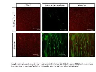
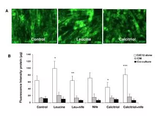
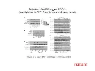
不飽和脂肪酸調控小鼠C2C12肌纖維細胞粒線體生合成之探討不飽和脂肪酸調控小鼠C2C12肌纖維細胞粒線體生合成之探討 • 近年來之動物及細胞實驗結果皆指出,當體內游離脂肪酸濃度增高時會促進肌肉中粒線體去偶合蛋白 (uncoupling protein; UCP) 表現,進而促進脂肪酸代謝。本研究旨在探討不飽和脂肪酸之n-9族油酸及n-3族亞麻油酸,對於肌肉細胞粒線體生合成相關基因、脂肪酸代謝相關基因及其上游轉錄因子基因表現之影響。以C2C12肌纖維細胞(C2C12 myotube)作為研究對象,投予0~1.2 mM之脂肪酸濃度進行24及48小時培養,觀察其細胞型態及檢測細胞存活速率。在測定基因表現方面,給予細胞0、0.3、0.6、0.9 mM之油酸及亞麻油酸培養24及48小時,再以同步定量反轉錄聚合?連鎖反應 (Real-time reverse-transcriptional polymerase chain reaction) 及西方墨點法,測定目標基因之表現。結果顯示,以0~1.2 mM油酸及亞麻油酸處理C2C12肌纖維細胞後,細胞型態於同一濃度及時間點時具一致性,但其細胞存活率則結果不同。經0.6~1.2 mM油酸處理24小時後,其細胞存活率為顯著降低之情形,但在48小時後則恢復與控制組相同;經0.3 mM亞麻油酸處理24小時後則會增加細胞存活率。以0.9 mM之油酸及亞麻油酸處理後皆會增加PGC-1α (peroxisome proliferators -activated receptor γ coactivator 1α) 之mRNA表現,且處理48小時後其蛋白質表現量之增加有濃度依賴性之現象。以0.6 mM油酸處理48小時後會伴隨增加COX I (cytochrome c oxidase subunit I) 之mRNA表現,而亞麻油酸則是以0.6~0.9 mM處理24小時即增加其粒線體flavoprotien及COX I mRNA表現。給予0.9 mM油酸及亞麻油酸處理48小時,皆會顯著增加其UCP3 (uncoupling protein 3)及CPT-1b (carnitine palmitoyl transferase-1b) 之mRNA表現。由上述實驗數據推論,油酸為經由促進PGC-1α之表現,亞麻油酸則並非完全經由PGC-1α途徑,來達到刺激粒線體生合成之效果;兩種脂肪酸皆會促進細胞內UCP3及CPT-1b之表現進而達到促進脂肪酸代謝之結果。
Modulation of Mitochondrial Biogenesis on C2C12 Myotube by unaturated Fatty Acids • Recently, uncoupling protein (UCP) located on mitochondrial inner membrane was found that its expression could be induced when plasma free fatty acids level increased to advance fatty acid metabolism. To evaluate effects of unsaturated fatty acids on mitochondrial biogenesis, mouse C2C12 myotubes were incubated with oleic acid (OA) or linoleic acid (LA). We observed cell morphology and analyzed cell viability after treatment with 0~1.2 mM fatty acid for 24 and 48 hr using phase contrast microscope and MTS (3-(4,5-dimethylthiazol-2-yl)-5-(3- carboxymethoxy -phenyl)-2-(4-sulfophenyl)-2H-tetrazolium, inner salt) assay. Expression of mitochondrial genes and related transcription factor genes were determined after treatment with 0~0.9 mM fatty acid for 24 and 48 hr using real-time RT-PCR and Western blot. Data showed that C2C12 myotube cell morphology in 0~1.2 mM OA and LA treatment were the same pattern in various time and concentration, but cell viability of OA and LA treatment were different. Cell viability was decreased in 0.6~1.2 mM OA treatment for 24 h, but it was recovered after 48 h treatment. After 0.3 mM LA treatment for 24 h would increase cell viability. Both OA and LA would induce PGC-1α mRNA and protein expression in C2C12 myotube with 0.9 mM treatment, especially after 48 h. 0.6 mM OA treatment would also induce COX I mRNA at 48 h. Besides, 0.6~0.9 mM LA treatment would induce mitochondrial genes including flavoprotein and COX I mRNA expression. Both OA and LA would induce UCP3 and CPT-1b mRNA expression with 0.9 mM treatment at 48 h. In conclusion, OA would stimulate mitochondrial biogenesis through PGC-1α pathway, but LA might stimulate by other way. Both OA and LA would promote fatty acid metabolism by increasing UCP3 and CPT-1b gene expression.