Wrist Trauma
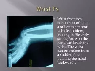
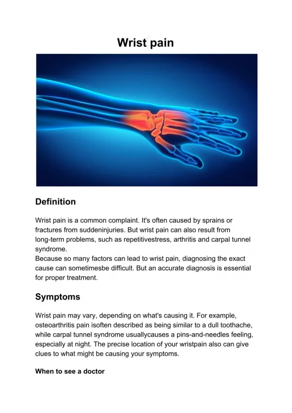
Wrist Trauma. Fractures and Dislocations of the Wrist. Clinically point tenderness over the wrist with >20% loss of grip strength are good physical indicators Complex anatomy requires four views for interpretation Neutral PA, PA in ulnar deviation, medial oblique and lateral

Wrist Trauma
E N D
Presentation Transcript
Fractures and Dislocations of the Wrist • Clinically point tenderness over the wrist with >20% loss of grip strength are good physical indicators • Complex anatomy requires four views for interpretation • Neutral PA, PA in ulnar deviation, medial oblique and lateral • Advanced imaging very useful because fractures not always visible
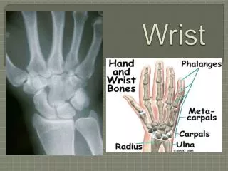
Normal Anatomy http://uwmsk.org/RadAnat/WristPALabelled.html
Distal Radius Fractures • Fractures may be subtle or even occult • Alteration of pronator quadratus fat plane is a useful indicator of fracture • Distal radial fractures include: -Colles’, Smith’s, Barton’s, Chauffer’s, Moore’s, torus, slipped radial epiphysis http://radiology.rsna.org/content/244/3/927.full
Distal Radius Fractures • Colles’ fracture- fx. of distal radius with posterior angulation of distal fragment • Usually FOOSH • Osteoporosis is risk factor, so increased incidence in women>65 • Smith's fracture (reverse Colles’ fracture)- fracture of distal radius with anterior angulation of distal fragment • Less common than Colles’ • Direct blow or fall on flexed hand http://imageinterpretation.co.uk/wrist.html
Distal Radius Fractures • Barton’s fracture (rim fx.)- posterior rim fracture of distal radial articular surface with associated proximal dislocation of carpals • Will see overlap of proximal row with articular surface of radius • Chauffeur’s fracture (backfire fx., Hutchinson's fx.)- fracture of radial styloid • Caused by avulsion or impaction by scaphoid • Formerly caused by starting cars with hand cranks http://www.radiologyassistant.nl/en/476a23436683b http://radiographics.rsna.org/content/24/4/1009/F11.expansion.html
Distal Radius Fractures • Moore’s fracture- fracture of ulnar styloid process and dislocation of distal ulna associated with Colles’ fracture • Torus fracture- buckling of cortex after trauma • Happens in children • Can happen in any long bone • Radiographic sign is bump or bulge of cortex
Distal Radius Fractures • Slipped radial epiphysis- childhood equivalent of Colles’ fracture • Hyperextension injury (FOOSH) causes shearing of radial epiphysis, which gets displaced posteriorly • Usually has small metaphyseal fragment (corner sign), which makes it Salter-Harris II http://www.oha.or.kr/skr/trauma/trauma4.htm
Distal Unlar Fractures • Ulnar styloid process fracture- rare as an isolated fracture • Usually avulsion by ulnar collateral ligament • More frequently found as associate fracture with other injuries • Distal ulnar shaft fracture- see “nightstick fracture” from forearm fracture section
Scaphoid Fractures • Most common carpal bone to fracture • Usually ages 15 to 40; rare in children • FOOSH • Clinical presentation is snuffbox pain with swelling • Most common site for occult fracture • Classified by anatomic location of fracture line • Waist (70%), proximal pole (20%) and distal pole (10%)
Scaphoid Fractures • Initially radiographs may be negative • Repeat x-ray in 10 to 20 days or advanced imaging immediately • MRI makes most sense if US not available • Complications include: • AVN, nonunion, carpal instability and radiocarpal degenerative arthritis
Scaphoid AVN • Scaphoid had 2 blood supplies • Proximal pole is supplied by an artery that enters distally • The more proximal the fracture is, the more likely AVN will occur if untreated • Radiographic signs include increased density (dead bone) and fragmentation
Nonunion • Missed diagnosis may lead to delay in treatment • Over time fracture line will widen and margins will become smooth and sclerotic
Carpal Instability • Follows rupture of scapholunate ligament • Radiographic features include: • Terry Thomas sign- widening of scapholunate jt. space >4 mm • Ring sign- rotation of scaphoid • Loss of parallel joint surfaces
Radiocarpal Degenerative Arthritis • Radiographic signs are that of OA anywhere else in the body • OA signs at the radiocarpal joint in the absence of trauma suggests diagnosis of CPPD • SLAC (ScaphoLunate Advanced Collapse) wrist may follow if capitate is allowed to migrate proximally
Triquetral Fractures • 2nd m.c. carpal bone to fracture • Usually by avulsion from dorsal surface by radiocarpal ligament (Fisher fracture) • Small flake dorsally only identifiable on lateral film
Fractures of Other Carpal Bones • All may fracture, but unusual unless directly traumatized • Hamate frequently complicated by non-union • Lunate frequentlyly complicated by avascular necrosis (Keinböck’s disease) http://orthoinfo.aaos.org/topic.cfm?topic=a00017
Dislocations of the Wrist • Two patterns • 1. a single bone that dislocates relative to remaining carpals • 2. a single bone that remains in place with the surrounding carpals dislocating • Evaluation of carpal arcs is a useful tool • Arc 1- proximal articular surfaces of proximal row • Arc 2- distal articular surfaces of proximal row • Arc 3- proximal surfaces of distal carpals (capitate and hamate) • Disruption indicates dislocation http://www.radiologyassistant.nl/en/42a29ec06b9e8
Single Carpal Dislocations • Lunate dislocation- most common carpal bone to dislocate • On PA film dislocated lunate appears triangular (pie sign) • Rows 2 and three disrupted • Scaphoid dislocation- • On PA film, see ring sign and Terry Thomas sign as previously described • Other single carpal dislocations unusual and require severe trauma
Multiple carpal dislocations • Perilunate dislocation- dorsal displacement of all carpals except lunate, which stays in place • On lateral, capitate does not sit in the lunate • On PA, capitate overlies lunate • Trans-scaphoid perilunate dislocation- same as above, but with associated scaphoid fracture • de Quervaine’s fracture dislocation- anterior dislocation of lunate as well as proximal fragment of fractured scaphoid http://www.imageinterpretation.co.uk/wrist.html
Fractures of 2nd - 5th Metacarpals • Boxers fracture- transverse fracture of neck of second or third metacarpals • Result of straight jab with fist • Barroom fracture- transverse fracture of neck of fourth or fifth metacarpals • Result of roundhouse blow from inexperienced fighter
First Metatarsal Fractures • Bennett's fracture- intra-articular fracture through base of the first metacarpal with dorsal displacement of the shaft • A small medial fragment remains at its articulation with trapezium • Rolando's fracture (comminuted Bennet’s) same as above but comminuted • Transverse fracture- most common fracture of first metacarpal • Doesn't interfere with articulation
Distal phalangeal fractures • Described as transverse, longitudinal, comminuted or chip fractures • Chip fractures occur at posterior or anterior corners of phalangeal base • Posterior chip fracture inactivates extension of the DIP joint and produces flexion deformity (mallet or baseball finger) • Middle phalangeal fractures • Chip fracture at anterior aspect of base (Volar plate fracture)
References Yochum, T.R. (2005) Yochum and Rowe’s Essentials of Skeletal Radiology, Third Edition. Lippincott, Williams and Wilkins: Baltimore.