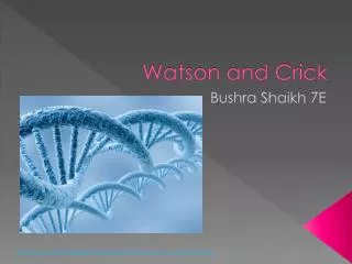
Figure 16.0 Watson and Crick
310 likes | 618 Views
Figure 16.0 Watson and Crick. Figure 16.0x James Watson. Figure 16.1 Transformation of bacteria. Figure 16.2a The Hershey-Chase experiment: phages. Figure 16.2ax Phages. Figure 16.2b The Hershey-Chase experiment. Figure 16.3 The structure of a DNA stand.

Figure 16.0 Watson and Crick
E N D
Presentation Transcript
Figure 16.4 Rosalind Franklin and her X-ray diffraction photo of DNA
Figure 16.7 A model for DNA replication: the basic concept (Layer 1)
Figure 16.7 A model for DNA replication: the basic concept (Layer 2)
Figure 16.7 A model for DNA replication: the basic concept (Layer 3)
Figure 16.7 A model for DNA replication: the basic concept (Layer 4)
Figure 16.9 The Meselson-Stahl experiment tested three models of DNA replication (Layer 1)
Figure 16.9 The Meselson-Stahl experiment tested three models of DNA replication (Layer 2)
Figure 16.9 The Meselson-Stahl experiment tested three models of DNA replication (Layer 3)
Figure 16.9 The Meselson-Stahl experiment tested three models of DNA replication (Layer 4)
Figure 16.11 Incorporation of a nucleotide into a DNA strand
Figure 16.13 Synthesis of leading and lagging strands during DNA replication
Figure 16.15 The main proteins of DNA replication and their functions
Figure 16.19a Telomeres and telomerase: Telomeres of mouse chromosomes