Download
1 / 50
510 likes | 707 Views
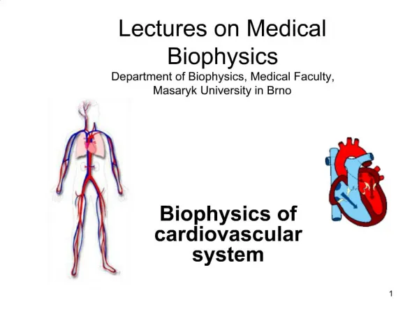
Biophysics of excitable cells. Axons are specialized for the conduction of an electrical impulse called an action potential. Specialized regions of neurons carry out different functions. Experimental techniques are conceptually simple. Cell-Semiconductor-Hybrids: Neuron on the Chip.

E N D
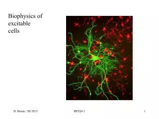
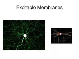
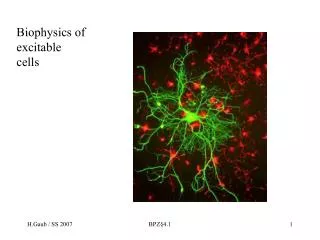
Biophysics of excitable cells BPZ§4.1
Axons are specialized for the conduction of an electrical impulse called an action potential BPZ§4.1
Specialized regions of neurons carry out different functions BPZ§4.1
Cell-Semiconductor-Hybrids: Neuron on the Chip More: Fromherz MPI Martinsried BPZ§4.1
Synapses are specialized sites where neurons communicate with other cells BPZ§4.1
Multiple exitatory and inhibitory synaptic contacts allow complex neuronal interconnects BPZ§4.1
Neurons are organized into circuits The knee-jerk reflex arc in the human. BPZ§4.1
Membrane depolarizations spread passively only short distances BPZ§4.1
The electrical activity of neurons results from the opening and closing of specific ion-channels proteins in the neuron plasma membrane BPZ§4.1
Voltage-gated cation channels generate action potentials BPZ§4.1
The structure and function of the voltage-gated Na+ channel BPZ§4.1
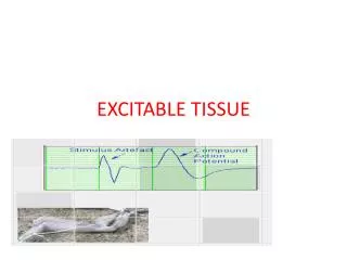
Action potentials are propagated unidirectionally without diminution Movements of only a few Na+ and K+ ions generate the action potential BPZ§4.1
Myelination increases the velocity of impulse conduction BPZ§4.1
Formation and structure of a myelin sheath in the peripheral nervous system BPZ§4.1
Each region of myelin formed by an individual glial cell is separated from the next region by an unmyelinated area called the node of Ranvier BPZ§4.1
Action potentials travel rapidly from one node to the next BPZ§4.1
Patch clamps permit measurement of ion movements through single channels BPZ§4.1
Current flux through individual voltage-gated channels determined by patch clamping of muscle cells BPZ§4.1
The oocyte expression assay can be used to determine if a protein is an ion channel BPZ§4.1
Voltage-gated K+ channels have four subunits each containing six transmembrane helices BPZ§4.1
All five subunits in the nicotinic acetylcholine receptor contribute to the ion channel BPZ§4.1
All pore-forming ion channels are similar in structure BPZ§4.1
Acetylcholine and other transmitters can activate multiple receptors Acetylcholine is released by motor neurons at neuromuscular junctions BPZ§4.1
Neurotransmitters are small molecules that transmit impulses at chemical synapses BPZ§4.1
Influx of Ca2+ triggers release of neurotransmitters Synaptic vesicles can be filled, exocytosed, and recycled within a minute BPZ§4.1
Synaptic-vesicle and plasma-membrane proteins important for vesicle docking and fusion BPZ§4.1
Ligand-gated receptor ion channels function at fast synapses BPZ§4.1
G protein-coupled receptors function at slow synapses BPZ§4.1
Transmitter-mediated signaling is terminated by several mechanisms • Following release of a neurotransmitter or neuropeptide, it must be removed or destroyed to prevent continued stimulation of the post-synaptic cell • To end the signaling, the transmitter may • diffuse away from the synaptic cleft • be taken up by the pre-synaptic neuron • be enzymatically degraded • Signaling by acetylcholine and neuropeptides is terminated by enzymatic degradation • Signaling by most classic neurotransmitters is terminated by uptake BPZ§4.1
Impulses transmitted across chemical synapses can be amplified and computed BPZ§4.1
Opening of acetylcholine-gated cation channels leads to muscle contraction BPZ§4.1
Cardiac muscarinic acetylcholine receptors activate a G protein that opens an ion channel Catecholamine receptors also induce changes in second-messenger levels that affect ion-channel activity BPZ§4.1
A serotonin receptor indirectly modulates K+ channel function by activating adenylate cyclase BPZ§4.1
Membrane disks in the outer segments of rod cells contain rhodopsin, a light-sensitive protein BPZ§4.1
Absorption of a photon triggers isomerization of retinal and activation of opsin BPZ§4.1
Cyclic GMP is a key transducing molecule in rod cells BPZ§4.1
A thousand different G protein-coupled receptors detect odors BPZ§4.1
Impulse transmission across electric synapses is nearly instantaneous BPZ§4.1
Comparison of action potential transmission across electric and chemical synapses BPZ§4.1
Learning and memory • Learning is the process by which animals modify their behavior as a result of experience or acquisition of information about the environment • Memory is the process by which this information is stored and retrieved • Long term memory involves the formation or elimination of certain synapses • Short-term memory involves changes in the release and function of neurotransmitters at specific synapses BPZ§4.1
Study of the gill withdrawal reflex of Aplysia has provided insight into short-term learning processes This simple behavior exhibits the most elementary forms of learning familiar in vertebrates: habituation, sensitization, and classical conditioning BPZ§4.1
Facilitator neurons mediate sensitization of Aplysia withdrawal reflex Individuals were restrained in small aquariums in a manner that the gill was exposed. A tactile stimulus was administered to the siphon and elicited the gill and siphon withdrawal reflex. A photocell was placed under the gill to record amplitude and duration of the response elicited by the stimulus. Habituation was observed when the stimulus was delivered repeatedly to the siphon. Stimulus every 90 seconds resulted in a rapidly declined response. By delivering an electric shock to the tail the response was rapidly restored, dishabituation occurred. Sensitization was observed when a strong stimulus was administered to the tail, this enhanced a completely rested reflex in Aplysia californica. BPZ§4.1
Coincidence detectors participate in classical conditioning and sensitization BPZ§4.1