Download
1 / 33
330 likes | 441 Views
Diagnostic Cytopathology & Significance of Biopsy Investigation. Jaroslava Dušková Inst. Pathol. ,1st Med. Faculty, Charles Univ. Prague https://www1.lf1.cuni.cz/~jdusk/. Morphological Diagnostic Methods. Clinical Pathological. Morphological Diagnostic Methods. Clinical:

E N D
Diagnostic Cytopathology & Significance of Biopsy Investigation Jaroslava Dušková Inst. Pathol. ,1st Med. Faculty, Charles Univ. Prague https://www1.lf1.cuni.cz/~jdusk/
Morphological Diagnostic Methods • Clinical • Pathological
Morphological Diagnostic Methods • Clinical: • macroscopy of lesions • visible with the naked eye • invisible with the naked eye - IMAGING (X-ray, sonography, scintigraphy, endoscopy, CT,…) • magnifying glass - colposcopy
Morphological Diagnostic Methods • Pathological • macroscopy • microscopy • ultrastructure • IMAGING
Morphological Diagnostic Methods • Pathological • macroscopy of lesions • autopsy report • biopsy description • cytology material description
Morphological Diagnostic Methods • Pathological • microscopy • cytology • minibiopsy - cytoblock • histology
Cytology (FNAB) • often both first and final dg. method • outpatient low cost procedure • done by an experienced (cyto)pathologist surprisingly effective • has some limits (!)
CYTOLOGY IS VERY EFFICIENT IN EARLY NEOPLASM DIAGNOSTICS
ESPECIALLY, IF CLINICO-MORPHOLOGICAL COMMUNICATION WORKS
Clinician from his pathologist: confirmation of neoplasm dg. nosological classification grading, staging prognosis reaction to the therapy recidive recognition Expectations
Pathologist from his clinician: information: local.,size, duration, former dg. a treated neo, clin. dg. diagnostic material acquisition correct interpretationof the pathologist´s report Expectations
GettingCytology Material • surface – smeared, brushed, scraped • cavities – punctured, aspirated • deep solid lesions - aspirated
ProcessingCytology Material • smears • cytospins, cytosedimentation • cytoblock
advantage of easy material taking together with more tissue architecture information histology &, immunohistochemistry methods available multilayered tissue phragments readable Minibiopsy - Cytoblock from FNAB
Cytology Material Staining • gynecology smears - polychrome • other materials –MGG, HE, polychrome, all other methods • cytoblock – multiple methods
Goals of Cytological Investigation • Screening – detection of symptomless lesions • Diagnosis of pathological lesions found • introductory (followed by histol.) • final
Gynecological oncologic cytology laboratory investigation standard Authors: MUDr Alena Beková, MIAC MUDr Pavel Tretiník, MIAC Oponents: doc. MUDr J. Dušková, CSc,FIAC MUDr Eva Svobodová
Cytology - Evaluation • staining and evaluation – minutes • Bethesda system: • material quality and quantity • group diagnosis • dg. as close to histology as possible • recommendation
Bethesda System 2001 <http://bethesda 2001.cancer.gov>
Suitable for Evaluation • without limits • limited by… • non processed • processed but limited for evaluation of squamous cell abnormalities due to…. .
General Categorisation • negative for intraepith. lesion or malignancy • intraepith. lesion or malignancy squamous or glandular • other pathology endometral cells in women over 40 yrs
Interpretation • negative for intraepithelial lesion or malignancy • Microorganisms • Trichomonas • mycosis vs. candidosis • shift - bact. vaginitis • bacteria Actinomyces like • cell changes of HSV type
Interpretation • negative for intraepithelial lesion or malignancy • Other non-neoplastic changes • reactive • inflammation (+ repair) • IUD • atrophy
Bethesda- cervical cytology classification 1. normal 2. benign cellular changes 3. ASCUS 4. L SIL 5. H SIL 6. Atypiae of glandular cells 7. susp. adenoca
Interpretation • epithelial abnormalities • squamous • ASC-US • ASC-H • LSIL CIN 1, HPV • HSIL CIN2, CIN3, CIS • susp. invasion • squamous ca
Interpretation • epithelial abnormalities • squamous • ASC-US • ASC-H • LSIL CIN 1, HPV • HSIL CIN2, CIN3, CIS • susp. invasion • squamous ca
Bethesda System 2001 <http://bethesda 2001.cancer.gov>
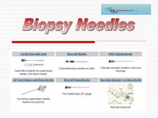
Cytology getting sample • needle 0.6-0.8mm • min. 2 punctions • aspiration • nonaspiration – reduction of the blood content • cyst: evacuate and aspirate with the second punction the periphery • fluid: wholevolume for cytology
Peroperation Biopsy • dg. during minutes • morphological artefacts (combination with cytology) • limited extensity of investigation • limited time
„Classical“ Biopsy (formol paraffin technique) • simple may be done in two days • immuno reactions two days more • further sectioning two days more • oncol. dg. - WHO classification • typing, grading, staging • prognostic factors
Biopsy • any tissue removed from a living patient is a COMPULSORY subject to • do not follow the tendency to discard „unimportant“ materials (nevi, tonsillae, uteri, intestine, endometrial curretage…..) !
Biopsy • do not crush small samples taking imprint cytology !!! • adequate amount of fixation solution (sample: fixative = min. 1:10) • wide neck bottle tightly closed for transport but openable without rough violence • flat sample – spread and tighten on a bearing socket • mark discrete suspicious lesion with a stitch before fixation
CYTOLOGYLOVE IT or LEAVE IT L. Cardozzo