Download
1 / 55
550 likes | 668 Views
Explore the intricate details of the cardiovascular system, including the heart's anatomy, blood flow, and cardiac muscle metabolism. Learn about important concepts such as heart valves, the cardiac cycle, and the electrical system governing heart function.

E N D
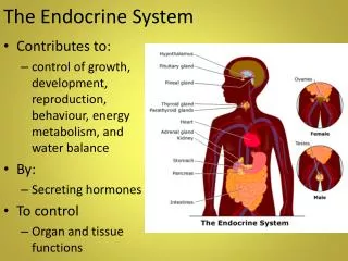
Cardiovascular System • Organ system which is responsible for the circulation of blood throughout the body • Consists of 2 main parts: • the heart contracts to move the blood vessels which: • Arteries transport oxygen rich blood away from the heart to the tissues. • allow for the exchange of substances between the blood and the cells of the body • Veins transport O2 poor CO2 rich blood back to the heart which is sent to the lungs to be oxygenated.
Heart • Considered to be a duel pump because the both sides work independently of one another. • the right side pumps blood to the lungs (pulmonary circulation) • the left side pumps blood to all of the other organs of the body (systemic circulation)
Heart Wall • Epicardium – Outermost layer ,visceral layer of the serous pericardium • Myocardium – cardiac muscle layer forming the bulk of the heart. • Endocardium – endothelial layer of the inner myocardial surface. • Deepest layer that is continuous with the lumen of the heart and arteries.
Coronary Arteries • Left coronary artery (LCA) • anterior interventricular branch • supplies blood to interventricular septum and anterior walls of ventricles • circumflex branch • passes around left side of heart in coronary sulcus, supplies left atrium and posterior wall of left ventricle • Right coronary artery (RCA) • right marginal branch • supplies lateral R atrium and ventricle • posterior interventricular branch • supplies posterior walls of ventricles
Heart Valves • Heart valves ensure unidirectional blood flow through the heart • Atrioventricular (AV) valves lie between the atria and the ventricles • Tricuspid Right • Bicuspid (Mitral) Left • AV valves prevent backflow into the atria when ventricles contract • Chordaetendineae anchor AV valves to papillary muscles • First heart sound (LUB) when valves close
During diastole there is less pressure in the in ventricle. AV Valves open and filling the ventricle. • During systole the AV valves prevent the back flow of blood into the atrium. • Failure to prevent the blood from going back into the atria (systolic heart murmur)
Heart Valves • Aortic semilunar valve lies between the left ventricle and the aorta • Pulmonary semilunar valve lies between the right ventricle and pulmonary trunk • Semilunar valves prevent backflow of blood into the ventricles • Second heart sound (DUB) when valves close
Figure 19.9b • During systole the Semilunar valves allow blood to be ejected from the ventricles into the aorta and pulmonary artery. • During diastole they prevent the back flow of blood back into the ventricles.
Cardiac Muscle Cell Metabolism • Aerobic respiration • Large mitochondria Rich in myoglobin and glycogen • Organic fuels: • fatty acids, glucose, ketones • Fatigue resistant
Anatomy of Heart Muscle • Cardiac muscle is striated, short, fat, branched, and interconnected • Intercalated discs anchor cardiac cells together and allow free passage of ions
Sequence of Cardiac Excitation • Note bundle branch and Purkinje fibers connection to both the ventricular myocardium and papillary muscles • This is important when considering how the ventricular myocardium and AV valves can function in a coordinated fashion.
Intrinsic Conduction System • Autorhythmic cells: cells that spontaneously generating action potentials without any influence from the CNS. • Main pacemaker of the heart is the (SA node) • sets cardiac heart rate. • Atrioventricular node (AV node) leads into Bundle of His which splits into branch bundles • Purkinjie fibers carry the impulse to the ventricular myocardium
Electrocardiography • Electrical activity is recorded by electrocardiogram (ECG) • P wave corresponds to depolarization of SA node • QRS complex corresponds to ventricular depolarization • T wave corresponds to ventricular repolarization • Atrial repolarization record is masked by the larger QRS complex
Cardiac Cycle • Cardiac cycle refers to all events associated with blood flow through the heart • Systole – contraction of heart muscle • Ejection on blood from the heart. • Diastole – relaxation of heart muscle • Myocytes repolarize allowing the heart to refill with blood • Coordination of the these phases is critical for proper cardiac function.
Phases of the Cardiac Cycle • Ventricular filling – mid-to-late diastole • At the beginning of the cycle, the semilunar valves are closed and the entire heart is in diastole. • Pressure in the heart blood is lower than in the vena cavas and pulmonary veins resulting in the movement of blood into the atria • As pressure in the atria now exceed ventricular pressure the AV valves are open allow blood to flow from the atria into the ventricles. • Atria systole occurs (SA) node initiates depolarization of both atria • Represented by the P-wave on an EKG • Both atrium contract forcing atrial blood into already filled ventricles. (End Diastolic Volume)
Phases of the Cardiac Cycle • Ventricular systole • Atria relax( repolarization) • There is a slight delay prior to the initiation of the AV node. • This allows atrial blood time to enter the ventricles • AV node sends action potential → along the bundle of His → branch bundles →perkinje fibers depolarizing both ventricles leading to the ventricle myocardium to contract. • QRS complex on EKG • This results in rising ventricular pressure causing the closing of AV valves (LUB) 1st heart sound • Isovolumetric contraction phase • (The ventricles are contracting with all 4 valves closed ) • This allows ventricular pressure to increase rapidly.
Phases of the Cardiac Cycle • Ventricular ejection phase: semilunar valves open once pressure in the ventricles exceed the pressure in the aorta and the pulmonary trunk. • Blood will now flow out of the heart.( Stroke Volume) • ST segment on EKG • Isovolumetric relaxation – early diastole • Ventricles relax represented by the T-wave on the EKG • Backflow of blood in aorta and pulmonary trunk closes semilunar valves making the second heart sound ( Dub) • This marks the end systolic volume (ESV) • Dicrotic notch – brief rise in aortic pressure caused by backflow of blood rebounding off semilunar valves • This rebounding of blood is when coronary artery perfusion takes place. ( the heart receives blood during diastole)
Cardiac Output • CO is how your body meets the demands that are placed on it. The heart must: • deliver vital nutrients such as O2, hormones and all the fuels sources body as quickly as they are used • remove CO2, urea, lactic acid… from the cells of the body as quickly as they are produced • Vigorous activities will increases systemic and cardiac O2 demands while producing more CO2 that will need to be expired • During strenuous activities the bodies CO will increase dramatically to meet these demands.
Cardiac Output (CO) • HR is the number of heart beats per minute • SV is the amount of blood pumped out by a ventricle with each beat • SV = end diastolic volume (EDV) minus end systolic volume (ESV) • EDV = amount of blood collected in a ventricle during diastole. • ESV = amount of blood remaining in a ventricle after contraction • CO is the amount of blood pumped by each ventricle in one minute • CO is the product of heart rate (HR) and stroke volume (SV)
Cardiac Output • Normal Cardiac Output • CO (ml/min) = HR (75 beats/min) x SV (70 ml/beat) • CO = 5250 ml/min (5.25 L/min) • Exercise CO can increase 7 fold • HR (160 beats/min) x SV (220 ml/beat) • CO = 35200 ml/min (35.25 L/min)
Extrinsic Innervation of the Heart • Heart is stimulated by the sympathetic cardioacceleratorycenter. • Sympathetic chain gangland from levels T1- T5. • Heart is inhibited by the parasympathetic cardioinhibitorycenter via the Vagas nerve.
Regulation of HR • The sympathetic cardiac nerve originating from the cardioacceleratory center in the medulla of the brain releases the neurotransmitter norepinepherine (adrenergic agent) onto the cells of the SA node • causes an increase in the frequency of action potentials in the SA node leading to an increase in HR. • What gates would you open to increase the frequency of action potentials? • Force of contraction is also increased because of the increased uptake of extra cellular calcium
Regulation of HR • Parasympathetic: Stimulation via (Vagus) nerve originating from the cardio inhibitory center in the medulla. • This center releases the neurotransmitter acetylcholine (cholinergic agent) onto the cells of the SA node • which causes a decrease in the frequency of action potentials in the SA node leading to a decrease in HR • Which gates will open?
Contractility and Norepinephrine • Sympathetic stimulation releases norepinephrine and initiates a cyclic AMP second-messenger system
Frank-Starling Law of the Heart • Preload : degree of myocardial stretch is related to the volume of blood in the ventricles .The greater the stretch on the ventricular walls, the greater the force the myocardium will contract thus increasing stroke volume. • Slower heart rate increase ventricular filling time (venous return) increasing SV • How will blood loss effect heart rate and stroke volume?
Factors Affecting Stroke Volume • Afterload – back pressure exerted by blood in the large arteries leaving the heart • The greater the afterload the harder the heart has to work to eject blood. • During diastole the coronary arteries fill with blood as blood recoils off the aortic semilunar valves. • Patients with chest pain (angina) are not getting enough blood to meet myocardial demand.
Blood Vessels • Blood is carried in a closed system of vessels that begins and ends at the heart • The three major, and types of vessels are arteries, capillaries and veins • Arteries carry blood away from the heart. • Veins carry blood toward the heart • 70% of blood is located in the veins • Capillaries contact tissue cells and directly serve as site for gas and nutrient exchange
Generalized Structure of Blood Vessels • Arteries and veins are composed of three tunics – • tunica interna, tunica media, and tunica externa • Lumen – central blood-fill area • Capillaries are composed of endothelium only: • necessary for exchange of gases and nutrients.
Tunics • Tunica interna (tunica intima) • Endothelial layer that lines the lumen of all vessels (inner most layer) • Tunica media (middle layer) • Smooth muscle and elastic fiber layer, regulated by sympathetic nervous system • Smaller vessels have a more muscular layer than larger vessels • SNS causes vasoconstriction. • (no stimulation ) results in vasodilatation of vessels • This will affect blood pressure and distribution) • Tunica externa (tunica adventitia) • Collagen fibers that protect and reinforce vessels • Larger vessels like the aorta have more to allow them to deal with higher pressures
Blood Flow Through Capillary • There are 5-6 liters of blood • 60 thousand miles of vessels in the circulatory system. • SNS causes precapillary sphincters to constrict shunting blood to where it is needed. • If all the precapillary sphincters open the blood pressure will drop dramatically. • This is what we call SHOCK.
Blood Pressure (BP) • Force per unit area exerted on the wall of a blood vessel. • Expressed in millimeters of mercury (mm Hg) • Systolic pressure – pressure exerted on arterial walls during ventricular contraction • Diastolic pressure – lowest level of arterial pressure during a ventricular cycle • The differences in BP within the vascular system provide the driving force that keeps blood moving from higher to lower pressure areas
Systemic Blood Pressure • The pumping action of the heart generates blood flow through the vessels along a pressure gradient, always moving from higher- to lower-pressure areas • Pressure results when flow is opposed by resistance • Systemic pressure: • Is highest in the aorta • Declines throughout the length of the pathway • Is 0 mm Hg in the right atrium • The steepest change in blood pressure occurs in the arterioles
Factors Aiding Venous Return • Venous BP alone is too low to promote adequate blood return and is aided by the: • Respiratory “pump” – pressure changes created during breathing suck blood toward the heart by squeezing local veins • Muscular “pump” – contraction of skeletal muscles “milk” blood toward the heart • Valves prevent backflow during venous return
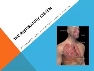
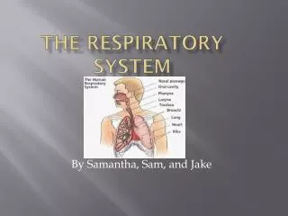
The Respiratory System Functions: • To provide the body with means of taking in(O2) for the production of ATP and eliminating (CO2) a byproduct of aerobic respiration. • To help maintain the body’s pH, by regulating the blood CO2 levels in the body. • Work in conjunction with the cardiovascular system to move these gases from the lungs to the cells and from the cells to the lungs.
Conducting Zone • Conducting zone • Provides rigid conduits for air to reach the sites of gas exchange • Respiratory structures include (nose, nasal cavity, pharynx, trachea, primary, secondary and tertiary bronchi) • No Gas exchange
Respiratory Zone • Respiratory zone: • begins as terminal bronchioles → respiratory bronchioles → alveolar ducts, → alveolar sacs composed of alveoli • This is where gas exchange occurs!
4 Processes of Respiration • Pulmonary ventilation – air moving into and out of the lungs along their pressure gradients. • Inspiration – air(O2)flows into the lungs • Expiration – air (CO2) exit the lungs • External respiration – gas exchange between the lungs (alveolus) and the blood (pulmonary capillaries) 3. Transport – transport of oxygen and carbon dioxide between the lungs and tissues via the circulatory system. • Internal respiration – gas exchange between systemic blood vessels (capillaries) and the tissues (cells) • Gases must diffuse into interstitial fluid prior to any exchange between the tissue and the cell.
Pulmonary Ventilation • Taking of air into and out of the lungs. • A mechanical process that depends on respiratory muscles changing the size of the thoracic cavity • Because this cavity is connected to the lungs via the parietal membranes it may also influence the lung (alveolar )volume. • A increase in alveolar volume will move air into the lungs down it concentration gradient. • A decrease in alveolus volume will move air out of the lungs.