Download
1 / 25
260 likes | 433 Views
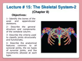
Lecture # 15: The Skeletal System-2. (Chapter 8). Objectives:. 1- Identify the bones of the axial and appendicular skeletons. 2- Describe the general structure and components of the vertebral column. 3- Describe the criteria used to classify joints structurally and functionally.

E N D
Lecture # 15: The Skeletal System-2 (Chapter 8) Objectives: 1- Identify the bones of the axial and appendicular skeletons. 2- Describe the general structure and components of the vertebral column. 3- Describe the criteria used to classify joints structurally and functionally. 4- Describe the anatomical features common to all synovial joints, the six types of synovial joints, and the movements allowed at each type.
The Appendicular Skeleton Upper limbs Pectoral girdle Pelvic girdle Lower limbs
Pectoral Girdle Right Clavicle Clavicles Superior view Scapulas Acromial end It articulates with the acromion of the scapula Sternal end It articulates with the manubrium of the sternum Acromial end Sternal end Anterior view Inferior view
Supraspionousfosa It articulates with the lateral end of the clavicle Acromion Spine Acromion Coracoid process Glenoid cavity Glenoid cavity Infraspionousfosa It articulates with the head of the humerus (a) Anterior view (b) Posterior view
Greater tubercle Head It articulates with the glenoid cavity of the scapula Olecranon Trochlear notch Intertubercular sulcus Lesser tubercle Coronoid process • An interosseus membrane unites the radio to the ulna. This joint is called syndesmosis Radius Ulna Coronoid fossa Olecranon fossa Capitulum Trochlea They articulate with the carpal bones Articular facets (a) Anterior view (b) Posterior view
Right Hand Carpal bones (8) Middle phalanx Proximal phalanx Metacarpal bones (5) I II V IV III Distal phalanx Phalanges (14) Metacarpal 1 Posterior view Distal phalanx Proximal phalanx
Right Coxal Bone Medial view Lateral view Iliac crest Ilium Ilium Greater sciatic notch Acetabulum Pubis Pubis Ischium Ischium Obturator foramen
Ilium Ischium Pubis Iliac crest Coxal bone Greater sciatic notch It articulates with the head of the femur Acetabulum Obturator foramen
Head Greater trochanter Greater trochanter It articulates with the acetabulum of coxal bone Femur Neck Lesser trochanter Linea aspera Posterior view Anterior view It articulates with the medial condyle of the tibia Medial condyle Patella Lateral condyle It articulates with the patella It articulates with the lateral condyle of the tibia Patellar surface
It articulates with the lateral condyle of the femur It articulates with the medial condyle of the femur Tibia Fibula • An interosseus membrane unites the fibula to tibia. This joint is called syndesmosis Lateral condyle Medial condyle Medial malleolus Lateral malleolus It articulates with the talus It articulates with the talus
Right Foot Superior view Lateral view Metatarsal bones (5) Tarsal bones (7) Calcaneus Talus Talus Phalanges (14) Metatarsal bones (5) V IV I III II Calcaneus First metatarsal bone Phalanges (14) Proximal phalanx Distal phalanx Proximal phalanx Middle phalanx Distal phalanx
Distal phalanx I Distal phalanx V Proximal phalanx I Middle phalanx V Proximal phalanx V Metatarsal II I III IV V Talus Calcaneus Key to tarsal bones Distal group Proximal group Superior (dorsal) view
JOINTS It is any point where two bones meet, whether or not the bones are movable at that interface Joint (articulation): Structural Classification (according to the manner in which the adjacent bones are bound to each other, with differences in how freely the bones can move) • 1- Fibrous joints: Points at which adjacent bones are bound by fibrous connective tissue (mainly collagen fibers). No space between the two bones • 2- Cartilaginous joints: When two bones are linked by cartilage. No space between the two bones • 3- Bony joints: Immovable joints formed when the gap between two bones ossify, and they become in effect, a single bone. No space between the two bones • 4- Synovial joints: They are joints in which two bones are separated by a space called a joint or synovial cavity Functional Classification (according to their functional characteristics or their ability to move) • 1- Synarthroses: Immovable joints • 2- Amphiarthroses: They are joints that allow slightly movements • 3- Diarthroses: They are freely movable joints
Structural Classification (according to the manner in which the adjacent bones are bound to each other, with differences in how freely the bones can move) • 1- Fibrous joints: Points at which adjacent bones are bound by fibrous connective tissue (mainly collagen fibers). No space between the two bones Sutures Gomphosis Syndesmosis Immovable or slightly mo-vable fibrous joints that closely bind the bones of the skull to each other • Collagen fibers attach tooth to jawbone allows the tooth • An interosseus membrane unites the fibula to the tibia (also the radio to the ulna)
2- Cartilaginous joints: When two bones are linked by cartilage. No space between the two bones • Bodies of vertebrae and intervertebral discs • Pubic symphysis in which right and left pubic bones joined by interpubic disc • 3- Bony joints: Immovable joints formed when the gap between two bones ossify, and they become in effect, a single bone. No space between the two bones Joints between the sacral vertebrae and bones of coccyx
4- Synovial joints: They are joints in which two bones are separated by a space called a joint or synovial cavity Ligaments They reinforce the joint and keep the bones in place Joint cavity or synovial cavity Joint capsule or articular capsule: Fibrous capsule It secrets a viscous and slippery synovial fluid that lubricates the joint Synovial membrane Articular cartilages They prevent friction between the bones
Functional Classification (according to their functional characteristics or their ability to move) • 1- Synarthroses: Immovable joints Gomphosis • 2- Amphiarthroses: They are joints that allow slightly movements Sutures • 3- Diarthroses: They are freely movable joints Synovial joints are diarthroses
The Six Types of Synovial Joints Ball-and-socket joint (humeroscapular) Hinge joint (humeroulnar) Pivot joint (radioulnar) Plane (gliding) joint (intercarpal) Saddle joint (trapeziometacarpal) Condylar joint (metacarpophalangeal)
The Six Types of Synovial Joints 1- Ball-and-Socket Joints 2- Hinge Joints • Smooth, hemispherical head fits within a cuplike socket. It is the only multiaxial joints in the body • Shoulder joint: head of humerus into glenoid cavity of scapula • Hip joint: head of femur into acetabulum of hip bone • One bone with convex surface that fits into a concave depression on other bone. It is mono- axial (move freely in one plane) • Elbow joint: ulna and humerus • Knee joint: femur and tibia • Finger and toe joints
3- Pivot Joints 4- Plane (gliding) Joints • One bone has a projection that is held in place by a ring-like ligament. One bone spins on its longitudinal axis. Monoaxial joint • Atlantoaxial joint: (dens of axis and atlas) • Proximal radioulnar joint: It allows the radius to rotate during pronation and supination • Flat articular surfaces in which bones slide over each other with relatively limited movement. Usually biaxial joint • Carpal bones of wrist • Tarsal bones of ankle
5- Condyloid (ellipsoid) Joints 6- Saddle Joints • Oval convex surface on one bone fits into a complementary shaped depression on the other. They are biaxial joints, movement in two planes • Radiocarpal joint of the wrist • Metacarpophalangeal joints at the bases of the fingers • Both bones have an articular surface that is shaped like a saddle, concave in one direction and convex in the other. It is more movable than a condyloid or hinge joint forming the primate opposable thumb. They are biaxial joints • Trapeziometacarpal joint (at the base of the thumb) Radio
Femur Femoropatellar joint - Patella Tibiofemoral joint Tibia The Knee Joint
Tendon of the quadriceps Synovial cavity Patellar ligament
Tendon of quadriceps collateral ligament collateral ligament patellar retinaculum patellar retinaculum Patellar ligament Quadriceps femoris muscle Fibular Tibial Lateral Medial
cruciate ligament meniscus cruciate ligament meniscus Tibial collateral ligament Lateral Medial Fibular collateral ligament condyle condyle Posterior Lateral Anterior Medial Patellar ligament Tendon of quadriceps