Download
1 / 28
470 likes | 1.25k Views
Surgical Disease of the Adrenal Gland (Part I). Roc McCarthy, D.O. Anatomy Physiology The incidental adrenal mass Fun facts Pheochromocytoma. Adrenal Gland. Cortex - mesoderm Medulla - neuroectoderm Renal agenesis, found in normal anatomic position Size- 5 x 3 x 1 x 5 cm

E N D
Surgical Disease of the Adrenal Gland (Part I) Roc McCarthy, D.O.
Anatomy • Physiology • The incidental adrenal mass • Fun facts • Pheochromocytoma
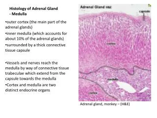
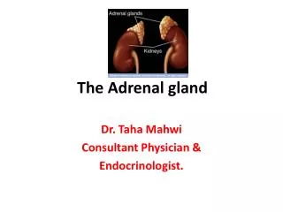
Adrenal Gland • Cortex - mesoderm • Medulla - neuroectoderm • Renal agenesis, found in normal anatomic position • Size- 5 x 3 x 1 x 5 cm • Retroperitoneal structure, contained in its own sub-compartment w/in Gerota’s fascia
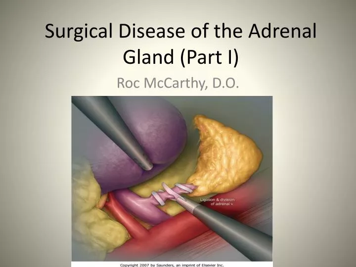
Adrenal Gland • Blood supply Arterial - receives 7cc/gram minute - 3 arterial sources of flow: 1. Inferior phrenic artery 2. Aorta 3. Renal artery Venous – single main vein most important surgical structure - right → post IVC - left → renal vein
Nerve Supply • Medulla - sympathetic branches - epinephrine - norepinephrine • Cortex – none • Lymphatics- lateral aortics (renal artery to diaphragm)
Aldosterone • Primary stimulus for release is angiotensin II • Other: ACTH, low serum Na, elevated K, JGA via low kidney perfusion
Adrenal Medulla • Distinct from cortex embryologically (neuroectoderm) • Secretes catecholamines sympathetic stimulation (mainly epinephrine but also norepinephrine and dopamine) • If pt. has excess of BOTH epi and norepi, the tumor is in the adrenal gland
The Incidental Adrenal Tumor • 0.5 – 5% of abdominal CTs show abnormal adrenal glands • 85% of adrenal masses are nonfunctional and BENIGN • Def. of incidental mass: - >1cm - discovered on exam for non-adrenal cause - absence of signs or symptoms of adrenal disorder
Questions to be asked: • Is the mass functional? • Any physical signs or symptoms? • Is there biochemical evidence of activity: 1. Pheo screen 2. Potassium level 3. Glucocorticoid screen • Is the mass malignant? • Any history of another malignancy? • Is imaging suggestive of malignancy?
Nature of Incidentally Found Adrenal Masses • Review of 2,005 incidentally-discovered adrenal masses: - Nonfunctioning adenoma 82% - Functioning: Cushing’s 5% Pheo 5% Aldosteronoma 1% - Malignancy: Metastasis 3% ACC 4% • Young WF, et al. Endocrinol Clin N Am. 2000
Good to Know Facts! • If the adrenal gland has fat density material, it is by definition a benign myelolipoma • If pt. has a known primary cancer, the adrenal mass with be mets from that site 50% of time • Overall 2-4% of adrenal masses are ACC • If mass >6cm, ACC 65%
Incidental Adrenal Mass Initial Evaluation • History and physical exam • Look for signs of hormonal syndromes • Search for occult malignancy • CXR • Stool for occult blood • Mammogram (women only)
Extent of Endocrine Evaluation? • Serum K (if HTN) → Conn’s • Plasma metanephrines: most sensitive test for pheo • 24-hr urine cortisol (Cushing’s)
Diagnosis: Imaging • Rare for a nonfunctional adenoma to become functional • MRI- Both ACC and pheo are hyper-intense in T2 images (light up from T1 to T2)
CT Adenoma Characteristics • Sharp margins • Smooth, homogenous, lipid rich • Most <10 Hu on noncontrast images • Washout >50% @ 15 min
Incidental Adrenal Mass Management • Hormonally active → surgical removal • > 5 cm → removal (with a scalpel) • < 3 cm observe • Surveillance Recommendations: Old: - CT at 6 months - Annual endocrine eval for 4 years New: If mass stable on scans @ 3m and 1 yr then no further workup
Pheochromocytoma • Incidence and Presentation - symptoms → release of epi/norepi - hypertension present 90% cases - orthostatic hypotension (low plasma vol) - 30% of pheo’s found at autopsy and cause of death cardiovascular disease - micturition syncope
Pheo (cont’d) • Triad: headache, tachycardia, diaphoresis • Other symptoms: Pallor, flushing, palpitations, abd/chest pain, weakness, N/V, psychosis • Small tumors more likely symptomatic • ALL patients regardless of age, have a complete cardiac work-up before surgery
Pheochromocytoma Rule’s of 10 • Bilateral • Familial (non-sporadic) • Pediatric • Malignant • Normotensive • Extra-adrenal • Multiple • *Childhood presentation breaks the rules- 25% bilateral, multiple, extra-adrenal
Pheo- The Diagnosis • Plasma free metanephrines- most sensitive test -seen 99% of patients • 24° urinary catecholamines (2x normal is diagnostic) • VMA • Clonidine suppression test (0.3mg oral, test 3 hrs later) >50% reduction catecholamines NO pheo
Pre-op Management • Early alpha blockade??? • Goal to control hypertension- phenoxybenzamine • Do NOT use b-blocker before alpha • IV hydration • Prevent cardiac arrhythmias
Pheo • Post-op - hypotension (most common) sec to secondary to hypovolemia • Surgical outcomes - excision does NOT always lead to long- term cure - recurrence 5% benign 10% malignant
Part II • Conn’s syndrome • Cushing’s syndrome • Addison’s disease • Adrenal cortical carcinoma • Metastatic disease to adrenal gland • Principles of adrenalectomy