Download
1 / 17
170 likes | 302 Views
This study explores the use of nanoscale silica particles for modifying surface topography to influence cellular responses, particularly in inhibiting cell proliferation. By applying cationic surfactant-coated silica on hydrophobic surfaces, the research demonstrates how nanoparticles can alter cellular adhesion and migration. The findings highlight the potential applications of this technology in wound dressings, hygienic surfaces, and medical implants. The results indicate that silica particles effectively inhibit fibroblast cell spreading and migration, regardless of protein concentration.

E N D
Surface modification using Nanoparticle to inhibit cellular proliferation B.G.Cousins, P.J.Doherty,, M.J.Garvey, R.L.Williams University of Liverpool U.K.
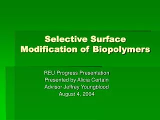
OPAL 1 micron
Diatoms Opal • The subunits of colloidal silica are non-porous, spherical particles of ~10nm • Silica is present in all connective tissues (collagen, arterial cell walls) along with nails, skin and hair • Exists in nature in plants, diatoms and opal
Colloidal Silica aggregates in aqueous dispersion to form 3-dimensional irreversible gels Silica Sol Silica Gel Irreversible Network Dissolution/ Deposition
Interface “Interfacial Gelation” of partially hydrophobed silica at an air or hydrophobic interface. Low levels of adsorbed cationic surfactant render colloidal silica surface active.
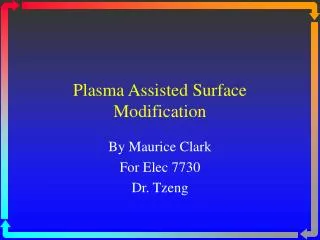
Two-dimensional aggregates silica 0.1 micron 2 micron
Air Water Silica Sol Monolayer Silica Sol Interfacial Gelation of partially hydrophobed silica at the air/water interface. LANGMUIR-BLODGETT Type Deposition
Electron micrograph of interfacial film of 14 nm silica particles
50 micron Interfacial rigidity of silica coating, observed at an oil/water interface Non-spherical oil droplets formed in cationic coated silica dispersion Rigid interface
MRC Discipline Hopper Award Deposition of Nanoparticles for Control of Cellular Response to Surfaces • Cells respond to nanoscale structures and adapt to topography • Deposition of nanoparticles offers a simple route to topographical modification. Potential applications in wound dressing, hygienic surfaces and implants.
Study Aim • The aim of the study was the manipulation of the surface topography to influence cellular response • Approach uses the deposition of commercially available, nanoscale, silica particles to modify the topography • Cationic surfactant coated silica limited to hydrophobic surfaces and necessitated an alternative deposition process.
ZetagTMPrimary Adhesive Layer N+R3 N+R3 N+R3 N+R3 N+R3 N+R3 N+R3 N+R3 N+R3 N+R3 N+R3 O- O- O- O- O- OH OH OH OH OH OH O- • Ionic adsorption of Zetag on to a glass surface is achieved via (O- N+R3) ion-pairs
SiO2 SiO2 SiO2 SiO2 SiO2 SiO2 SiO2 SiO2 + + + + + + + + Deposition of Silica • Negative silica particles adsorb on to the cationic polymer
Results • 21 nm Silica particles attached to glass substrate • Image is representative of 7, 14, 21 and 80 nm silica particles
Core technology Intraocular lenses Wound dressings Surface Modification Using Nanotechnology Bone fracture repair devices Potential Applications of Core Technology
Plain glass Plain glass Nanoparticles Nanoparticles Cell Response to Nanotopography • Fibroblast cells on coated and uncoated glass • Silica particles inhibit adhesion/spreading of cells • Silica particles prevent migration of cells • This effect is not altered by changing protein concentration • Effect seen with other cell types (primary & established)