Understanding the Body's Function: Anatomy and Movement in Physical Activity
This course explores the four major systems of the human body—skeletal, muscular, respiratory, and cardiovascular—and their roles in physical activity performance. Students will gain knowledge about the axial and appendicular skeletons, the structure and function of muscles, and the types of joints. Through examining how these systems work together, learners will better appreciate the anatomy's contribution to movement and coordination, essential for any physical exercise or sport.

Understanding the Body's Function: Anatomy and Movement in Physical Activity
E N D
Presentation Transcript
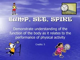
BUMP, SET, SPIKEDemonstrate understanding of the function of the bodyas it relates to the performance of physical activity Credits: 5
Functional Anatomy The body has 4 Major Systems: - Skeletal System – the bone structure for support and protection of the organs - Muscular System – muscles connect to bones to allow movement - Respiratory System – lungs which take oxygen from the lungs and transfers it to the blood - Cardiovascular System – heart and veins/arteries which pump blood for energy production
AXIAL SKELETON Provides a framework that supports and protects the organs in the body. Skull:Protects the brain and guards the entrance to the digestive and respiratory systems Ribs:Protects the heart and lungs, and helps with the function of breathing Vertebral Column:Provides a column of support to protect the spinal cord and keep us in an upright position
THE SPINE 7 Cervical Vertebrae 12 Thoracic Vertebrae 5 Lumbar Vertebrae – Main weight bearing bones 5 Fused Vertebrae (Tailbone)
THE RIBCAGE 12 Pairs of Ribs: 1-7 = True Ribs 8-12 = False Ribs 8-10 Join to the cartilage 11-12 are Floating Ribs
APPENDICULAR SKELETON • Includes the bones of the arms and legs and the joints that connect them to the Axial Skeleton. • It gives you control over your environment, changes your position in space and provides mobility
APPENDICULAR SKELETON The bones of the Upper and Lower Limbs
UPPER LIMBS Clavicle: For attachment of many muscles Scapular: Shoulder blade for protection of the pectoral girdle (shoulder joint) Humerus: Main weight bearing bone in the arm for lifting objects Ulna: The smaller forearm bone which extends from the pinky. The top of the bone that attaches to the Humerus is shaped like a U Radius: The larger forearm bone which helps in support and rotation of the forearm Carpals: The 8 bones of the wrist which allow finer movement Metacarpals: The 5 bones of the hand Phalanges: The 14 finger bones. The thumb consists of 2 phalanges while each finger has 3 phalanges.
LOWER LIMBS Pelvis: For protecting the reproductive organs and sustain all the internal organs Femur: Main weight bearing bone in the body Patella: For protecting the knee joint. It is a sesamoidbone as it is not attached to any other bones Tibia: (On Top) The shinbone for transfer of weight through the ground Fibula: The smaller of the two bones for attachment of muscles to move the foot and toes Tarsals: Bones of the ankle to allow for flexion when walking Metatarsals: 5 bones of the foot for balance and walking Phalanges: 14 bones of the toes
JOINING BONES In order to keep our body together we need a number of connective tissues to be the glue for our body. LIGAMENTS - these are a band of tissues which connects Bone to Bone. cartilage - this is a pad of fibrous tissue which separates bones or provides a cushion to prevent rubbing.
MUSCULAR SYSTEM • Muscles attach to the bone to allow movement to occur. They are in charge of pulling on bones in order to create movement. There are approximately 639 skeletal muscles in the body. Muscles need a different type of tissue to attach to the bone – it must be capable of withstanding tension (like a rubber band). This tissue is called a Tendon.
DIRECTIONS OF THE BODY SUPERIOR - Top POSTERIOR - back ANTERIOR - front INFERIOR - Bottom
UPPER BODY - BACK Trapezius: Neck muscle Deltoid: Shoulder muscle Tricep: Muscle at the back of the arm Latissimus Dorsi: Wing (back) muscles Flexor Carpi: Forearm muscles
UPPER BODY - FRONT Trapezius Pectorals: Chest muscles Deltoid Abdominals: Core (trunk) muscles Obliques: Side muscles Extensor Carpi: Forearm muscles
LOWER BODY - BACK Gluteus Maximus: Bottom muscles Hamstrings: Back of the thigh Gastrocnemius: Calf muscle Soleus: Ankle muscle
LOWER BODY - FRONT Quadracep: Thigh muscle Tibialis Anterior: Shin muscle
JOINTS There are 3 types of Joints: 1. Immovable Joints 2. Slightly Movable Joints 3. Freely Movable Joints
Immovable Joints: These are joints that cannot move. They are extremely strong joints where the bones are either interlocked (like a zip) or they are fused. • These joints are found in the head. They are: • between the bones of the skull • between the teeth and jaw
Slightly Movable Joints: These are joints that have a small amount of movement. They are connected by either ligaments or cartilage. Ligaments attach bone to bone. One example is between the Tibia and Fibula. Cartilage is a pad of fibrous tissue. It separates and provides a cushion for joining bones. It is found between the vertebrae and the two pubic bones
Movable Joints: These are joints that have movement in one (i.e. elbow) two (i.e. wrist) or three (i.e. shoulder) different planes or directions. These joints are also known as: SYNOVIAL JOINTS
SYNOVIAL JOINTS Articular Cartilage:Cover the bony surfaces so the bones cannot touch each other. It is slick and smooth to reduce friction during movement Joint Cavity: The gap between the two bones which contains the synovial fluid Synovial Fluid: Found in the joint cavity, it is a fluid that has three purposes: 1. Lubrication – to reduce friction when moving 2. Nutrient Distribution – moves nutrients around and collects waste 3. Shock Absorption – acts as a cushion when the joint is squashed
TYPES OF SYNOVIAL JOINTS • Multiaxial – slide across a surface in many directions Gliding Joint – they have a flat surface where one bone can slide across the surface of another. The movement is only very small. e.g. Sacroiliac Joint (Between the sacrum and the pelvis) Intercarpal Joints (Between the carpal (wrist) bones) Claviculosternal Joints (Between the clavicle and the sternum)
TYPES OF SYNOVIAL JOINTS • Monoaxial – Joints that move in one direction Hinge Joint – allow a bending motion like the opening and closing of a door e.g. Elbow Joint Ankle Joint Phalangeal Joints (fingers) Knee Joint Pivot Joint – only allow rotation (turning) of a joint e.g. Atlas/Axis Vertebrae
TYPES OF SYNOVIAL JOINTS • Biaxial – Joints that move in two directions Saddle Joint – Shaped like a horses saddle that can move back and forth and side to side. e.g. Thumb Joint Ellipsoidal Joint – Shaped like a herb/pesto bowl that can move back and forth and side to side. e.g. Finger Joints Wrist Joint
TYPES OF SYNOVIAL JOINTS • Triaxial – Joints that move in three directions Ball and Socket – Shaped like and egg in a cup, they can move back and forth, side to side, and round-and-round e.g. Shoulder Joint Hip Joint
DESCRIBING MOVEMENT • Movement is described by looking at the way the joints are positioned when a movement occurs. • The main joints involved in movement are the Shoulder, Elbow, and Wrist (in the upper body), and the Hip, Knee, and ankle (in the lower body). When the muscle pull on the bones that surround these joints, movement occurs.
TYPES OF MOVEMENT – Angular Motion FLEXION – to reduce the angle between the two bones EXTENSION – to increase the angle between the two bones HYPEREXTENSION – to increase the angle further than the normal position
TYPES OF MOVEMENT – Angular Motion ABDUCTION(ab, from)– To move away from the longitudinal axis of the body (normal standing position) ADDUCTION(ad, to)– to move back toward the normal position
TYPES OF MOVEMENT – Angular Motion CIRCUMDUCTION – to draw a large circle
TYPES OF MOVEMENT – Rotational Motion ROTATION – to turn around an axis. It may be: • Left or Right • Inward or Outward
TYPES OF MOVEMENT – Rotational Motion PRONATION – turns the wrist and hand from palm facing front to palm facing back SUPINATION – turns the wrist and hand from palm facing back to palm facing front
TYPES OF MOVEMENT – Special Movement INVERSION – to twist the foot inward (side to side) EVERSION – to twist the foot outward
TYPES OF MOVEMENT – Special Movement DORSIFLEXION – to lift the foot and flex at the ankle joint i.e. when you dig your heel PLANTARFLEXION – to point your foot and extend your ankle joint i.e. plant your foot on the accelerator
SKELETAL SYSTEM - Practice Skull Humerus Clavicle Sternum Ulna Scapula Radius Carpals Ribs Metacarpals Phalanges Vertebrae Pelvis Femur Sacrum Patella Tarsals Tibia Metatarsals Fibula Phalanges
MUSCULAR SYSTEM - Practice 1 7 2 8 9 3 10 4 11 5 6
MUSCULAR SYSTEM Deltoids Trapezius Tricep Pectoral Bicep Latissimus Dorsi Abdominals Gluteus Maximus Quadricep Hamstring Gastrocnemius
MOVEMENT AT THE JOINTS • When we break movements down, we can think about which movement happens at each Joint. • We can also think about how many different directions they can travel in. • These directions include: Direction 1: Forward and Backward Direction 2: Side to Side Direction 3: Around (circle or rotation)
MOVEMENT • Forward and Backward movement is known as Flexion and Extension • Side to side movement is known as Abduction and Adduction • Around movement is known as Circumduction OR Rotation.
MUSCLE PAIRS • When muscles create movement they must work in pairs for this to happen. Impulses from nerves cause muscles fibers to contract (muscle contraction). This causes the muscles to become shorter and thicker. This causes the bone to move. • For this to happen, the opposite muscle must relax to allow for this movement. This causes this muscle to become longer and thinner. • For the muscle to return to its normal position, these pairs swap over.
The muscle contracting and causing the movement is called the Agonist. The relaxed muscle that allows the movement to occur is called the Antagonist.