Download
1 / 32
320 likes | 481 Views
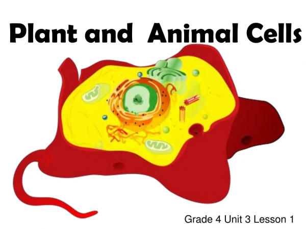
Ch. 3 Cells!. Introduction. Cell theory Cells are the smallest living subunit of an organism All cells arise from pre-existing cells Bacteria, amoebas are unicellular and function independently Human cells work interdependently Vary in size, shape, function Mostly microscopic

E N D
Introduction • Cell theory • Cells are the smallest living subunit of an organism • All cells arise from pre-existing cells • Bacteria, amoebas are unicellular and function independently • Human cells work interdependently • Vary in size, shape, function • Mostly microscopic • More than 200 kinds
Cell Structure • Cell membrane (plasma membrane) • Forms outer boundary of cell • Nucleus • Absent in mature red blood cells • Cytoplasm • Organelles
Cell Membrane • Plasma membrane (PM) • Made of phospholipids, cholesterol, proteins (Fig. 3-1) • Phospholipidbilayer • Permit lipid-soluble materials to enter/leave cell via diffusion • Cholesterol makes membrane more stable • Proteins • Form channels or pores to permit passage of materials • Carrier enzymes (transporters) help substances enter cell • Antigens identify cells as “self” • Receptor sites for hormones
Cell Membrane (cont.) • Active, dynamic membrane • Selectively permeable • Certain substances can pass through, others cannot
Nucleus • Within cytoplasm • Bounded by 2-layered, porous nuclear membrane • Contains 1 or more nucleoli, chromosomes (Fig. 3-2) • Nucleolus – small sphere of DNA, RNA, protein • Form ribosomal RNA (rRNA); becomes part of ribosomes; involved in protein synthesis
Nucleus (cont.) • Control center • Contains 46 chromosomes in long threads called chromatin • Before cell division, chromatin coils into visible chromosomes (made of DNA, protein) • Nucleus contains the same genetic information, but only a small number of genes are active “switched on” • Active genes code for proteins
Cytoplasm • Watery solution of minerals, gases, organic molecules, organelles • Cytosol • Water portion of cytoplasm; where many rxns take place
Organelles • Intracellular structures, often membrane-bound • Have specific functions in cell metabolism (Fig. 3-2) • Endoplasmic reticulum (ER) • Extensive tubular network extending from nuclear membrane to PM • Rough ER has ribosomes on surface, smooth ER does not • Passageway for proteins, lipids
Organelles (cont.) • Ribosomes • Not membrane-bound • Very small structures made of protein and rRNA • Some on Rough ER, others in cytoplasm • Site of protein synthesis
Organelles (cont.) • Golgi apparatus (GA) • Flat, membranous sacs stacked on top one another • Carbohydrate synthesis, packaging for secretion from the cell
Organelles (cont.) • Golgi Apparatus Secretion • Small sacs of Golgi membrane break off & fuse with PM • Substance is released to cell exterior (exocytosis)
Organelles (cont.) • Mitochondria • Oval or spherical organelles bounded by double membrane • Inner membrane has folds (cristae) • Site of ATP production • Contain own genes in a single DNA molecule • Duplicate themselves when cell divides • Many mitochondria in muscle cells
Organelles (cont.) • Lysosomes • Single-membrane digestive structures • Have enzymes that digest bacteria, old cell parts and dead cells
Organelles (cont.) • Centrioles • Pair of rod-shaped structures just outside the nucleus • Organize spindle fibers during cell division • Cilia • Short, hair-like structures covering surface of cell • Beat in unison and sweep materials across cell’s surface • Cilia in fallopian tubes sweep egg toward the uterus • Flagella • Whip-like tail • Provides motility for sperm
Organelles (cont.) • Microvilli • Folds of cell membrane on the free surface of a cell • Greatly increase surface area of membrane • Part of cells lining organs that absorb material • Small intestine • Kidney tubules • Table 3-1
Cellular Transport Mechanisms • Enable cells to move materials into or out of cell • Diffusion • Osmosis • Facilitated diffusion • Active transport • Filtration • Phagocytosis • Pinocytosis
Diffusion • Movement of molecules from an area of high to low concentration along a concentration gradient • Oxygen and CO2 move by diffusion (Fig. 3-3)
Osmosis • Diffusion of H2O through a selectively permeable membrane • Water moves from an area with more to an area with less water • Water will move to an area with more solutes • If a 2% salt solution and a 6% salt solution are separated by a membrane, allowing only water to pass through, in which direction will water diffuse? • See Box 3-1
Facilitated Diffusion • Molecules move through a membrane from an area of high to low concentration with assistance • Use a carrier enzyme (transporter)
Active Transport • Requires ATP to move molecules from a low to high concentration against a concentration gradient • Nerve and muscle cells have sodium pumps to move sodium ions out of cells (Fig. 3-3)
Filtration • Requires energy of mechanical pressure • Water, dissolved materials are forced through a membrane from an area of high to low pressure • Formation of tissue fluid; first step in urine formation
Phagocytosis & Pinocytosis • Forms of endocytosis • Phagocytosis • White blood cell engulfs bacteria (Fig. 3-3) • Digestion using enzymes in lysosomes • Pinocytosis • Stationary cells take in small molecules adsorbed or attached to their membranes • Kidney tubules reabsorb small proteins (Fig. 3-3) • Table 3-2
Cell Division • Process by which cell reproduces itself • 2 types • Mitosis • Meiosis
Mitosis • One cell with a diploid (2N) # of chromosomes divides into 2 identical cells, each with the diploid number • 2N = 46 • Interphase • DNA replication enables each chromosome (chromatin) to copy itself • Resting (non-dividing) stage • Stores energy in ATP
Mitosis (cont.) • Long, thin, invisible chromatin begins to coil • Each looks like a letter X because original DNA molecule & its copy (chromatids) are attached • Mitosis stages (PMAT); see Table 3-4; Fig. 3-5 • Prophase • Metaphase • Anaphase • Telophase
Mitosis (cont.) • Essential to replace damaged or dead cells • Occurs constantly in epidermis, stomach lining, red bone marrow • Does not occur in most muscle cells & neurons • Skeletal muscle cells have limited mitosis • Research has found some potential for mitosis in the CNS and heart • At present, mitosis does not take place sufficiently enough to replace dead cells
Meiosis • More complex • Results in gamete (egg, sperm) formation • One diploid cell (2N) divides twice to form 4 haploid cells (N) • Meiosis takes place in ovaries (oogenesis) and testes (spermatogenesis) • “reduction division” • During fertilization, the egg joins with the sperm to restore the diploid number of 46 in the fertilized egg (zygote)


![[virtual] cells](https://cdn1.slideserve.com/3553683/slide1-dt.jpg)