Download

1 / 41

E N D
Prevalent as early as ancient Greek times. Hippocrates described the cutaneous spreading of lesions. Shakespeare is thought to have been familiar with these lesions and their transmission and mentioned in his Romeo and Juliet. In 1893 Vidal recognized the human transmission of HSV infection from one individual to another. In 1919, Lowenstein confirmed experimentally the infectious nature of HSV. In 1920's and 1930's, the natural history and range of infections of HSV were studied. By the 1940's and 1950's, research established on diseases caused by HSV. HERPES VIRIDAE
Herpesviridae, a large family of DNA viruses that cause diseases in animals and humans. The members of this family are also known as herpesviruses. Name is derived from the Greek word herpein ("to creep or crawl"). Latent, reactivation , recurring and lytic infections are typical of this group of viruses.
Medically important viruses – Three subfamilies. Alpha herpes virinae. Rapid growth, Latent infection in sensory ganglia. HSV – 1, HSV – 2, V – Z Virus. Beta herpes virinae Slow growth. Grow best in Fibroblasts. Latent infection in salivary gland. HHV – 5 ( CMV ) HHV– 6 , HHV – 7
Gamma herpes virinae Grow in Lymphoblastoid cells. Latent infection in Lymphoid tissue. HHV – 4 (Epstein – Barr Virus) HHV – 8 ( Kaposi’s sarcoma herpes virus) Neurotropic viruses: HSV,VZV; Lymphotropic viruses: EBV,HHV6,HHV7
Morphology Enveloped (Lipid) Double stranded DNA Icosahedral capsid “Tegument” Glycoprotein spikes ( Surface spikes) Naked virus
Replication and susceptibility Virus replicates in host cell nucleus. Cowdry type Aintranuclear inclusion bodies. Susceptible to Ether Chloroform Bile salts. Heat labile.
1.Continuous cell line cultures. Monkey or Rabbit kidney. Human amnion cell line cultures. HeLa cell cultures. Cytopathic effects : Well defined foci with heaped up cells & syncitial formation.
2.Growth on Chick embryo CAM Shiny, non necrotic pocks.
Trigeminal, Sacral Physical, Emotional stress. Trauma, Fever. Sun light. Pathogenesis Enters ‘thru’ defects in skin &mucus membranes. Local multiplication Local LN involvement. Retrograde axonal flow from sensory nerves …. Reaches Ganglia. Maintains latency Viral replication in the nerves. Decreased CMI. Centrifugal migration to skin & mucus membranes. Recurrence of the disease.
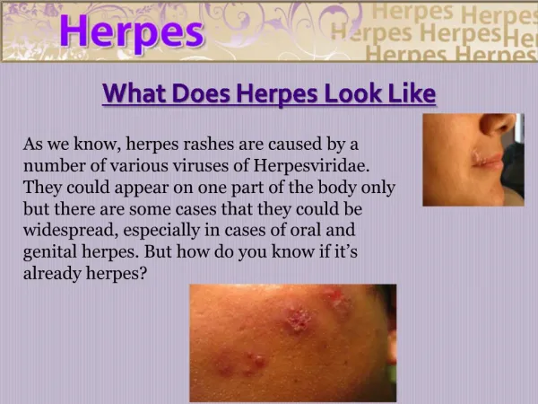
Primary infection Vesicle formation (Ballooning degeneration of intra-epithelial cells) Site of eruption shows Pain, tingling, warmth & itch, erythema & papule Thin walled umbilicated vesicleroof breaks down and forms an ulcer. with fever (cold sore or fever blisters). Mucus membrane, non-keratinized epithelia. Recurrent infections are seen with severe pain, ulceration.
Neuroinvasiveness • Neurotoxicity • Latency in dorsal root ganglion of CNS • Damaged epithelium is repaired by Natural • killer cells. • Viral glycoproteins initiates the T-cell • activity which activate primed B cells to • produce antibodies.
Pathological lesions Herpetic gingivostomatitis 1. Mucocutaneous infections Cheeks, chin, forehead. Napkin rash in infants. Acute gingivostomatitis Around the mouth & on lips. Pre-school children. Majority of primary infections; Teething / oral thrush. Ruptured lesions
Herpetic lesions in oral cavity Dental anomalies followed by Herpes viral infections. .
Skin infections Herpetic whitlow , Toddlers, Dentists, Nurses, Herpes gladiatorum (Wrestlers) Painful, swollen, grouped vesicles with pus.
Eye infections: Branching/dendritic corneal ulcer. Eczema herpeticum Severe form of Atopic eczema(Kaposi's varicelliform eruption.) Extensive ulceration.
Visceral HSV: Esophagitis Tracheobronchitis Pneumonitis CNS: Sporadic, fatal encephalitis(HSV 1). Seizures, Hemi paresis and paraesthesia Congenital infections : Transplacental infection. Congenital malformations are rare.
HSV- 2 transmitted byauto infection, sexual /orogenital contact. Most infections are asymptomatic. -Symptomatic infections with sores, fever and lymphadenopathy of genital tract, heal within 2-4 weeks. -Patients may suffer with 4-5 outbreaks (recurrent) within a year. -Causes psychological distress. -Infection during late pregnancy poses a greater risk of transmission to the baby. -Sacral radiculopathy common with urinary retention.
Genital herpes Perianal herpes Labial herpes Shaft of the Penis
2.Neonatal herpes Localized to Skin , eyes, mouth. Disseminated infection leads to Multi organ involvement (Liver, Adrenals, Brain) Complication: Neurological impairment. 3.Aseptic meningitis:
Laboratory diagnosis Microscopy: Tzanck smear 1 % Aqu. sol. of Toludineblue. Multinucleated giant cells with faceted nuclei , ground glass chromatin. (Tzanck cells) Best : Giemsa, Papanicolou stain 1. Smears Scrapings from base of Vesicle CSF, Saliva. Serum -Primary infection. -ELISA most useful. -CFT.
Treatment :No cure. Acyclovir : Primary infection (< 72 hrs). Reduce the recurrences. Famciclovir: For resistant cases. 3.Antigen detection : Fluorescent antibody , ELISA 4.Viral isolation: Cell line culture – growth within 1 -3 days. 5.Molecular techniques: PCR and DNA probes.
Varicella-Zoster virus Varicella Mildest childhood exanthemata Source: Patient Droplet nuclei from Respiratory tract Inhalation. Incubation period 7– 23 days Centripetal distribution. Macule Papule Vesicle Pustule Scab Vesicular rash surrounded by a ring (Trunk) Superficial “Drop of water”
Appears in crops, Profuse in adults Hemorrhagic & bullous. Interstetial Pneumonia. Postviral encephalitis. Guillain- Barre syndrome. Recovery is spontaneous. Can cross placenta Neuromuscular disorders.
Lab. Diagnosis : Same as HSV. Specimens : Buccal / Cutaneous lesions Prophylaxis : V – Z immunoglobulins. Live attenuated varicella vaccine.
Herpes Zoster (Creeping girdle) Herpes zoster ophthalmicus. Old age (>60 years). Latent virus in dorsal root or cranial nerve ganglia Neuritic pain,Parasthesiafor weeks / months. Unilateral, painful eruption in thoracic region. Commonest sites: Areas innervated by spinal cord segments D3 – L2 & Trigeminal nerve.
Complications : LMN Paralysis. Meningo-encephalitis Ramsay Hunt syndrome Facial palsy + eruption on tympanic membrane & external auditory canal
Cytomegalovirus (Salivary gland virus) Infected cells : Cytomegaly (Owl’s eye appearance) Most of the infections are unapparent. Commonest cause for congenital defects. Pathogenesis: Latent infection : Mononuclear leucocytes (Monocytes, B -lymphocytes) Secretary glands Kidney. Replication seen in ductal epithelial cells. Excretion in body fluids, Milk & Urine.
Modes of transmission Transplacental. Sexual contact. Blood and its products. Organ transplantation. Urine, Saliva, Cervical secretions, semen, breast milk. Perinatal & Postnatal infections: Infected birth canal,Breast milk.
1.Transplacental route: Condition severe if infection occurs during first trimester of pregnancy. Cytomegalic Inclusion disease of Newborn(10%) characterised by varied type of clinical manifestations. Hepatospleenomegaly Jaundice Thrombocytopenic purpura Haemolytic anaemia Microcephaly Cerebral calcifications. Mental retardation.
IMN like disease: Young adults. Hepatitis, fever, atypical lymphocytosis. (No pharyngitis, no lymphadenopathy, Negative for heterophile antibody) Respiratory tract infections: Pneumonitis in infants. Immunocompromised individuals: Pneumonia Fatal encephalitis Chorioretinitis
Lab. Diagnosis: Adults:Urine, Saliva, BAL, Semen & Cervical secretions. Neonate: Urine 1. Microscopy: Centrifuged deposits of secretions. Giemsa stain: Cytomegalic cells
2.Isolation: “Human diploid fibroblast cell” culture. Large retractile cells with cytoplasmic granules. 3.Serology: Anti CMV IgM Ab estimation by ELISA. Treatment: Ganciclovir & Foscarnet. Prophylaxis: Acyclovir.
Epstein – Barr (EB) Virus Burkitt's lymphoma in 1964 . Affinity for B – lymphocytes (CD 21 receptors.) 80 – 90% of children by three years of age. Asymptomatic. Not highly contagious. Droplets are not infectious. Source : Saliva, Oropharyngeal secretions
Pathogenesis Infected saliva. Pharyngeal epithelial cells (Multiplies locally).Persistence Shed in saliva Invades blood stream Infects B. lymphocytes. Liver Spleen Polyclonal activation cell death . Unchecked replication results in Lymphomas Neoantigen formation Atypical lymphocytosis.
Clinical Diseases Incubation period : 4 – 7 weeks 1.Infectious Mononucleosis (Glandular disease, Kissing disease) Acute self limiting illness. Fever, sore throat. Lymphadenopathy. Sub clinical Hepatitis, Tender spleenomegaly Abnormal lymphocytes in PS
2.Chronic fatigue syndrome. 3.Malignancies associated with EB. Burkitt’s lymphoma (Malignant B cell lymphoma of jaw) Nasopharyngeal carcinoma Lymphomas in HIV infected persons.
Lab diagnosis 1.Blood smear examination :Atypical Lymphocytosis. 2.Paul - Bunnel test: Heterophile antibody detection test. Inactivated serum + 1% sheep RBC suspension 370C 4 hrs Agglutination (>100) 3. EBV Specific antibodies: EBNA Ab EBNA Ig M VCA, Ig G VCA 4. PCR: More sensitive.
HHV – 6 Isolated in 1986 from AIDS patient T - lymphocytotropic (CD+) Transmission through Oral secretions. Roseola infantum (Exanthema subitum) High fever with generalized rash. Chronic fatigue syndrome.
HHV – 8 Kaposi's sarcoma related herpes virus. In 1994 : Association with Kaposi's sarcoma. (Rare type of “B cell lymphoma” from AIDS patients). HHV – 7 Isolated from AIDS patient in 1990. No disease association. Remains as orphan virus. 39
Research is currently ongoing into a variety of side-effect or co-conditions related to the herpesviruses. Alzheimer's disease atherosclerosis cholangiocarcinoma Crohn's disease chronic fatigue syndrome fibromyalgia Irritable bowel syndrome multiple sclerosis pancreatic cancer pityriasis rosea Type II Diabetes
Prepared fore-learningby Dr .P.SRINIVASULU REDDY , M D., Professor, Department of Microbiology Narayana Medical College NELLORE 41