Download
1 / 29
290 likes | 495 Views
Topic 4: Movement Analysis. Neuromuscular function Muscle contraction. How does the muscular system contribute to human movement?. Nervous system connecting with muscular system.

E N D
Topic 4: Movement Analysis Neuromuscular functionMuscle contraction How does the muscular system contribute to human movement?
Nervous system connecting with muscular system • The central nervous system (CNS) consists of brain and spinal cord and is where most sensing and control takes place. • Sensory neurons carry signals to the CNS from receptors that sense various factors such as body temperature, blood pressure, blood oxygen and carbon dioxide levels (and more). =>Afferent neurons • Motorneurons are nerves which carry information from the CNS to the muscles and which tell muscles to contract or relax. => Efferent neurons
Skeletal muscle has important functions • Movement: skeletal muscle attach to bones, against which they pull to enable movement • Support and Posture: the muscles are seldom fully relaxed and are often in a constant state of slight contraction. This is known as muscle tone • Heat production: the contraction of skeletal muscle involves the production of energy. In breaking down glycogen to provide this energy heat is released. This is why the body gets hot while exercising
(Taken from: Sport & PE, Students book: A complete guide to Advanced Level Study)The structure of Skeletal Muscle The muscle belly is surrounded by a layer of epimysium(1.15a) a thick connective tissue surrounding the entire surface of the muscle. This is continuous and eventually forms tendons which join the muscle onto bones. The muscle belly is composed of many bundles of fibers know as fasciculi, these are covered by the perimysium. Each fiber within a single fascicle contains many smaller fibers called myofibrils(1.15b) which provide the contractile unit. Myofibrils have characteristic dark and light bands (striations) which represent a sarcomere (1.15c). This pattern is repeated along the length of the myofibril Prefixes sarco and myo both refer to muscle. Therefore if you see a word with either of these prefixes you should immediately think MUSCLE.
So, does muscle contract by itself? • No, for skeletal muscle to contract it needs an interaction with the nervous system (neuromuscular interaction). When a muscle is required to contract, an electrical impulse is emitted from the central nervous system. The electrical impulse, or action potential, begins at the brain and is transmitted via the spinal cord and by nerve cells called motor neurons. One motor neurone(nerve) cannot stimulate the whole muscle, but is only capable of stimulating a numbers of fibers within it. The motor neurone and the muscle fibers it stimulates, is called a motor unit.
Motor units • A motor unit is a motor neuron and all of the muscle cells (muscle fibers) it stimulates. The strength of a muscle contraction is determined by the size and number of motor units being stimulated. • The number of muscle fibers innervated by a single motorneuron may be high as 2000 or as small as 10. • Why the difference in the innervation ratio?
Diagram of motor unit www.tokresource.org
Motor units allow for selective contraction of muscle fibers so that we may control the strength and extent of muscle contraction. Without motor units a nerve impulse to the muscle would result in the entire muscle contracting to its full extent. That would make every motion that we make an “all or none” motion. This type of movement would make life nearly impossible. This is why the muscle fibers are surrounded by a membrane called the endomysium, This is very important in the physiology of muscle contraction because it electrically insulates the individual muscle cells from each other. • At the ends of the muscle all of the connective tissue sheaths (epimysium, perimysium, and endomysium) converge to form a tendon which will connect the muscle to its attachment site.
Role of neurotransmitters in human movement • Neurotransmitter: A chemical that is released from a nerve cell which thereby transmits an impulse from a nerve cell to another nerve, muscle, organ, or other tissue. A neurotransmitter is a messenger of neurologic information from one cell to another. (http://www.medterms.com) • Neurotransmitters like Acetylcholine are the chemicals which allow the transmission of signals from one neuron to the next across synapses. They are also found at the axon endings of motor neurons at the motor end plate, where they stimulate the muscle fibers. • Acetylcholine/cholinestrase in the neuromuscular junction • Neuromuscular junction • Muscle contraction
The structure of Actin and Myosin • Sarcomeres have a highly organized structure, and at the most fundamental level the sarcomere is composed of two protein-based myofilaments: • A thick myosin filament • A thinner actin filament • The interaction and overlapping of these two myofilaments enables muscles to contract through the sliding filament theory • http://www.youtube.com/watch?v=Ct8AbZn_A8A • Skeletal muscle (Sarcomere, myosin and Actin)
Summary • Electrical signal from CNS triggers a release of acetylcholine in the synapse -> Influx of Sodium ions and efflux of potassium ions reach a threshold for an action potential -> This action potential travels along the myofibril over the sarcolemma (through the T-tubules) and causes the sarcoplasmic reticulum underneath to release calcium ions (Ca2+) into the sarcomeres -> Sarcomeres are composed of two protein-based myofilaments, actin (thin) and myosin (thick) The Calcium ions opens binding sites on tropomyosin (string of protein) connected to troponin coiled around actin filaments. The myosin head connects to the actin and using the energy from a chemical called ATP (adenosine triphosphate) that is on the myosin heads it bends myosin head “sliding” actin along (sliding filament theory) -> When the energy is released to cock the head of myosin ATP is broken into ADP (adenosine diphosphate) and phosphate. Nerve no longer stimulated, an enzyme called acetylcholinesterase breaks down the acetycholine and it returns to the synaptic vesicles.
Control of muscle force Done by recruiting motor units in two ways: • Size principle – smaller motor units are recruited first, larger motor units later when larger forces are required. • Frequency (rate coding) of motor unit recruitment – higher rate of activation of motor units causing higher force in muscle
Measuring muscle activation • To measure the electrical activation of a muscle in an athlete, detremining the timing of contraction, force of contraction and fatigue, is useful. • This is done with Electromyography (EMG), using electrodes in or on the surface of the muscle. • Is this invasive/accurate? Raw EMG signal (action potential in muscle) and rectified signal.
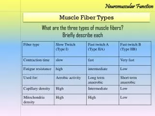
Fast twitch & slow twitch muscles • Type 1 – • Type 2a – • Type 2b - Slow twitch, slow transmission speeds, small muscle forces, fatigue resistant (red fibers) Fast twitch, fast contractions, stronger contractions, fatigue resistant (pink, Fast.Oxidative.Glycolitic. fibers) Fastest contractions, largest forces Fatigue quick (white, F.T.G. fibers)
Major differences in structure and function between slow- and fast twitch muscle fibers • Speed of contraction – slow-twitch muscle fibers contract at a rate of about 20% when compared with fast-twitch muscle fibers • Muscle fiber force – fast-twitch fibers are bigger in size than slow-twitch fibers, have larger motor neurones and therefore can generate high force rapidly • Muscle endurance – slow-twitch fibers are capable of resisting fatigue whereas fast-twitch are easily fatigued • Mitochondrial density – Slow-twitch (and type 2a) muscle fibers have higher mitochondrial density compared to fast-twitch muscle fibers coloring the muscle red. Fast-twitch fibers are white. (type 2a is pink) • Glycogen stores – Fast-twitch muscle fibers have far bigger glycogen stores than slow-twitch
Taken from Sport and PE, A complete guide to advanced level study.
Glycogen General characteristics • Polysaccharide (C6,H10,O5) • Stored primarily in the liver and muscle tissue • Readily converted to glucose needed by the body to satisfy energy needs • Supplies energy during heavy work • Stored with water (1gr Carb with 3gr Water) • CNS is dependent on hepatic (from liver) glycogen Effects on performance • Increased storage can double duration of exhaustive work • Low or depleted glycogen stores - limits exercise intensity - decreases time to exhaustion -increases rating of perceived exhaustion during physical activity http://www.exrx.net/Nutrition/Glycogen.html http://student.biology.arizona.edu/honors99/group7/glycogen.jpg
Which muscle fibers have a higher glycogen content? Fast twitch and slow twitch muscle fibers
http://www.abc.net.au/science/news/img/health/womensprintA300904.jpghttp://www.abc.net.au/science/news/img/health/womensprintA300904.jpg
http://www.anytimeroswell.com/images/iStock_000003059041XSmall.jpghttp://www.anytimeroswell.com/images/iStock_000003059041XSmall.jpg
http://www.nervemedia.org.uk/wp-content/uploads/2011/09/Ironman_World_Triathlon_Championship5.jpghttp://www.nervemedia.org.uk/wp-content/uploads/2011/09/Ironman_World_Triathlon_Championship5.jpg
http://www.chinadaily.com.cn/english/doc/2005-11/12/xin_2411021217017383196725.jpghttp://www.chinadaily.com.cn/english/doc/2005-11/12/xin_2411021217017383196725.jpg
http://cdn.bleacherreport.net/images_root/slides/photos/001/404/968/kelechi-osemele_display_image.jpg?1318232343http://cdn.bleacherreport.net/images_root/slides/photos/001/404/968/kelechi-osemele_display_image.jpg?1318232343
http://1.bp.blogspot.com/-bJCLQ_uExes/TZbwWpsqEOI/AAAAAAAAAV0/kmWebVnyc18/s400/table-tennis-players1.jpghttp://1.bp.blogspot.com/-bJCLQ_uExes/TZbwWpsqEOI/AAAAAAAAAV0/kmWebVnyc18/s400/table-tennis-players1.jpg
http://cdn.bleacherreport.net/images_root/slides/photos/003/105/872/hi-res-156942461_crop_650.jpg?1366037949http://cdn.bleacherreport.net/images_root/slides/photos/003/105/872/hi-res-156942461_crop_650.jpg?1366037949