Download
1 / 95
950 likes | 987 Views
Family Medicine: Gait Analysis, Knee Injuries, Foot/Ankle Injuries and Overuse Injuries. Selina Silva, MD Associate Professor, Pediatric Orthopaedics Medical Director, UNM Carrie Tingley Hospital. Outline. Gait Analysis Knee injuries Foot/Ankle injuries Pediatric overuse injuries.

E N D
Family Medicine: Gait Analysis, Knee Injuries, Foot/Ankle Injuries and Overuse Injuries Selina Silva, MD Associate Professor, Pediatric Orthopaedics Medical Director, UNM Carrie Tingley Hospital
Outline • Gait Analysis • Knee injuries • Foot/Ankle injuries • Pediatric overuse injuries
Gait Analysis • Can be informal with clinical evaluation of gait during a routine visit • Tip: have child walk in hallway if they are older. Easier to see abnormalities. • Can also get formal gait analysis in a gait laboratory.
Gait lab • Video analysis of children walking to aid in preoperative planning. • Goal would also be for every child with movement disorder (cerebral palsy, muscular dystrophy, etc) have a simple video of them walking stored annually. • Research is another large parts of plans for the gait lab
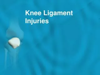
Knee Injuries • Anatomy and Physical Exam • ACL • MCL • PCL • Patella Dislocations
Anatomy • ACL • PCL • MCL • LCL • Meniscus • Medial • Lateral
Inspection Palpation Range of Motion Special tests Neurovascular assessment Physical Exam of the Knee
Effusion Erythema Ecchymosis Edema Q angle Angular deformities Muscular asymmetry INSPECTION
ANTERIOR Tibial tubercle Infrapatellar tendon Quad insertion Patellar facets Crepitus ? MEDIAL MCL Meniscus Pes anserine insertion Tibial plateau Femoral condyle PALPATION
ACL Special Tests • Anterior drawer • Lachman test • Pivot shift test • Valgus stress test at full extension!
ACL: PHYSICAL EXAM • Decreased ROM • Effusion-hemarthrosis, immediate • + Instability tests • Lachman: most accurate • Pivot shift • Anterior drawer • + MCL and meniscus tests
LIGAMENT INJURIES: XRAY • AP • Lateral capsular sign: Segond fx • Tibial spine avulsion fx • Physeal injuries • Lateral • Lateral condyle divot • Obliques ? • Tangential (Merchant) Lateral capsular disruption
ACL TREATMENT • Grade 3 Injuries- Surgery • Indications • Most active people will require surgery to restore adequate function and decrease instability • Recurrent instability • Inability to modify activity • Associated injuries: meniscus • Age? • Wait three weeks due to arthrofibrosis risk • 100% @ 6-12 months
MCL INJURIES HISTORY • Mechanism = valgus stress • Medial joint line pain • Lack of large effusion • Difficulty weight-bearing
MCL Injuries - Physical Exam • Tender to palpation along MCL • Pain + instability with valgus stress • 30o flexion = MCL • 90o flexion = associated ACL • COMPARE SIDES
MCL Injuries – Treatment Grade 1,2 • Early mobilization • Weight-bearing as tolerated • Hinged knee brace • Recovery 4-6 weeks
MCL Injuries – Treatment Grade 3 • Isolated = nonsurgical management • Combined = surgery consistent with associated injuries • Natural Hx = lack of long-term degenerative changes seen with ACL, meniscus
PCL INJURIES • Mechanism • Sports = fall on flexed knee with foot plantarflexed, hyperextension, pivot • MVA = dashboard injury • Effusion (less than with ACL) • Shifting/instability (chronic) • Less distinctive
PCL Injuries – Physical Exam • + Effusion • + Posterior drawer test • + Posterior sag sign • False positive Lachman test • Common to have isolated injuries
PCL Injuries - Treatment • Functional bracing (early) • Rehab • Surgery if continued instability, effusions • Note- 2% of NFL preseason exam with incidental isolated PCL tear
PATELLAR INSTABILITY • Acute patellar dislocation • Acute patellar subluxation • Patellar tracking dysfunction
PATELLAR DISLOCATION History • Mechanism = pivot • Immediate effusion • May visualize patella dislocated laterally • + Instability (chronically) Frequently Patella spontaneously relocates
Patellar Dislocation – Physical Exam • Tender peripatellar structures • Medial retinaculum • Lateral femoral condyle • Effusion • ? Patella dislocated laterally X-rays- osteochondral fracture, effusion
PATELLAR DISLOCATION Treatment • Knee extension immobilizer in ER • Early quad setting exercises • Change to Shield’s Brace • Return to sport • Full, painless ROM • Normal strength • Adequate aerobic fitness
Fractures and Dislocations about the Knee in Pediatric Patients
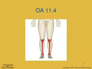
Anatomy • Distal femoral physis- large, undulating- irregular • Proximal tibial physis- contiguous with tibial tubercle apophysis • Ligament and muscular attachments may lead to avulsion injuries, fracture angulation
Anatomy- Neurologic and Vascular Structures • Popliteal artery tethered above and below knee • Common peroneal nerve vulnerable at fibular neck/head
Distal Femoral Physeal Fractures • Direct blow mechanism • Salter I or II common • Check neurologic & vascular status
Distal Femoral Physeal Fractures • Closed reduction and pinning for displaced fractures • Long leg cast
Distal Femoral Physeal Fractures • High rate of premature growth arrest • rare < 2 yo • 80% 2 - 11 yo • 50% > 11 yo • Angular deformity • Leg length discrepancy
Patella Fractures in Children • Largest sesamoid bone, gives extensor mechanism improved lever arm • Uncommon fracture in skeletally immature patients • May have bipartite (superolateral) patella- avoid misdiagnosis
Patellar Sleeve Fracture • 8-12 year old • Inferior pole sleeve of cartilage may displace • May have small ossified portion • <2mm displaced, intact extensor mechanism- treat non-operatively
Patella Sleeve Fractures • Avulsion mechanism • Management same as adults • Restore articular surface and knee extensor mechanism
Tibial Tubercle Fractures • Primary insertion of patellar tendon into secondary ossification center of proximal tibia • Mechanism- jumping or landing, quadriceps resisted contraction • Common just before completion of growth (around 15 years in males)