Terminal Bronchioles
190 likes | 368 Views
Terminal Bronchioles. Lined by simple ciliated columnar epithelium without goblet cells No cartilages, glands. Respiratory Bronchioles. Branches of terminal bronchioles Give rise to alveolar sacs, alveolar ducts, and alveoli Not sites of actual gas exchange

Terminal Bronchioles
E N D
Presentation Transcript
Terminal Bronchioles • Lined by simple ciliated columnar epithelium without goblet cells • No cartilages, glands
Respiratory Bronchioles • Branches of terminal bronchioles • Give rise to alveolar sacs, alveolar ducts, and alveoli • Not sites of actual gas exchange • Lining epithelium: simple cuboidal, non-ciliated epithelium • Some Clara cells
Alveolar Ducts • Branch of from the walls of respiratory bronchioles • Conical, thin-walled tubes • Lined by simple squamous epithelium • Give rise to numerous alveolar sacs and alveoli that the openings practically occupy the entire wall of the duct
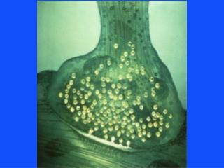
Alveolar Sacs and Alveoli • Sites of actual exchange of gases between blood and inspired air • Arise from either respiratory bronchioles or alveolar ducts • Thin-walled polyhedral sacs • Open on one side – allow entry of air from the respiratory bronchioles or alveolar ducts
Alveolar Sacs and Alveoli • Around 300 million • Packed so closely that each alveolus does not have a separate wall • Adjacent alveoli share a common wall (interalveolar septum)
Interalveolar Septum • Consists of thin supporting layer of connective tissue covered on either side by simple squamous epithelia • Alveolar pores– allow direct communication between the cavities of adjacent alveoli
Interalveolar Cells • Type I (pulmonary epithelial cell, small alveolar cell, pneumonocyte type I cell) • Type II (great alveolar cell, pneumonocyte type II cell)
Type I Interalveolar Cell • Less numerous but cover a majority of the alveolar surface area (> 95%) • Squamous or flattened cells • Form the structure of the alveolar wall
Type II Interalveolar Cell • Secrete pulmonary surfactant– reduces alveolar surface tension, thus prevents collapse of alveoli at the end of expiration • Cuboidal or round cells
Pulmonary Alveolar Macrophages • “Dust cells” • Most numerous cells in alveoli • Float freely in alveoli • Phagocytes
Blood-Air Barrier • Ultra-thin structure separating the blood in a pulmonary capillary from the air in the alveolus • Consists of: • Pulmonary epithelial cell • Basal lamina of the alveolar epithelium • Basal lamina of the capillary endothelium • Capillary endothelial cell