Download
1 / 10
110 likes | 359 Views
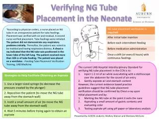
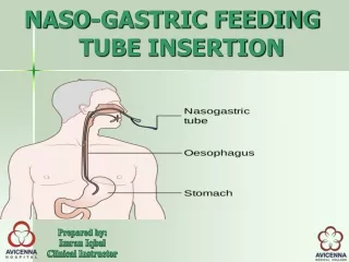
Verification of Feeding Tube Placement (blindly inserted). Issued August 2010. Expected Practice. Use a variety of methods to predict location during tube insertion Signs of respiratory distress Capnography if available Visual characteristics of aspiration

E N D
Verification of Feeding Tube Placement(blindly inserted) Issued August 2010 Verification of Feeding Tube Placement
Expected Practice • Use a variety of methods to predict location during tube insertion • Signs of respiratory distress • Capnography if available • Visual characteristics of aspiration • Ausculatory and water bubbling are unreliable Verification of Feeding Tube Placement
Expected Practice • Obtain radiographic confirmation of any blindly inserted tube • Radiograph should visualize the entire course of the tube • Should be read by a radiologist • Mark and document the tube’s exit site immediately after confirmation of correct placement Verification of Feeding Tube Placement
Expected Practice • Check tube location at 4 hour intervals after feeding is started • Observe for change in length of the external portion of the tube • Review routine chest and abdominal x-rays for tube location • Measure pH of aspirates • Observe appearance of feeding tube aspirates • If there is doubt about placement – obtain an x-ray Verification of Feeding Tube Placement
Scope and Impact • Blind placement of a feeding tube can cause serious and even fatal complications. • Even a small percentage of such complications can affect a significant number of people. • Styleted small-bore tubes are most often associated with complications, large-bore unstyleted tubes are not without Nasogastric feeding tubes were malpositioned in 1.3% to 2.4% of all insertions, malpositions resulted in pneumonia. • Critically ill patients often have multiple risk factors for airway misplacements; among these are a decreased level of consciousness, altered gag reflex, presence of an endotracheal tube, and multiple insertion feeding tubes may be malpositioned in the brain. • Risk for aspiration is greatly increased when a feeding tube’s ports end in the esophagus. • Complications related to malpositioned feeding tubes can be minimized by explicit policies and procedures for feeding tube insertions. Verification of Feeding Tube Placement
Bedside Methods to Determine Placement • Signs of respiratory distress • Capnography • pH and Appearance of Aspirate • Listening over the epigastrum for air insulfflated through tube Verification of Feeding Tube Placement
Radiographic Confirmation • Properly obtained and interpreted radiograph is recommended • Marking and documenting the tube at exit after confirmation of correct placement Verification of Feeding Tube Placement
Checking Tube Location at Regular Intervals • Change in length of external tube • Review routine chest and abdominal x-rays • Testing PH of feeding and appearance of tube aspirate • Listening over epigastrum for air insufflated through tube • Obtain x-ray tube location if in doubt Verification of Feeding Tube Placement
Actions for Nursing Practice • Use a variety of techniques to assess tube placement during insertion • Obtain x-ray that visualizes entire course of newly inserted tube • Ensure that your unit has written policies and procedures • If not already in place; develop documentation practices • Monitor tube position at 4 hour intervals Verification of Feeding Tube Placement
Need More Information? For more information or further assistance, please contact a clinical practice specialist with the AACN Practice Resource Network. Email: practice@aacn.org Verification of Feeding Tube Placement